Clear Sky Science · en
State-dependent neuronal and network dynamics in the lateral hypothalamus across sevoflurane anesthesia–emergence revealed by microelectrode arrays
How sleep-like brain states help anesthesia work
Anyone who has gone under general anesthesia has felt its strange switch-like power: one moment you are awake, the next you awaken with no memory of what happened. This study peeks under the hood of that switch in the mouse brain, focusing on a deep region called the lateral hypothalamus that helps keep us awake or asleep. By watching individual brain cells and larger brain waves at the same time, the researchers show how this area changes activity as animals move from wakefulness into sevoflurane anesthesia and then back to consciousness.
Listening in on a tiny wakefulness hub
The lateral hypothalamus is packed with different kinds of cells that either promote wakefulness or stabilize sleep. It also talks to many other crucial regions involved in arousal and motivation. To follow what these cells do during anesthesia, the team engineered ultra-thin microelectrode arrays, or MEAs, that can be gently slipped into this deep brain area of mice. They coated the tiny metal contacts with a special mix of platinum particles and a conducting polymer to lower electrical resistance and boost signal quality. Tests showed that this surface treatment sharply reduced noise and allowed the electrodes to record robust neural signals for weeks, laying the technical foundation for stable tracking of brain activity.
A three-part journey: awake, under, and back again
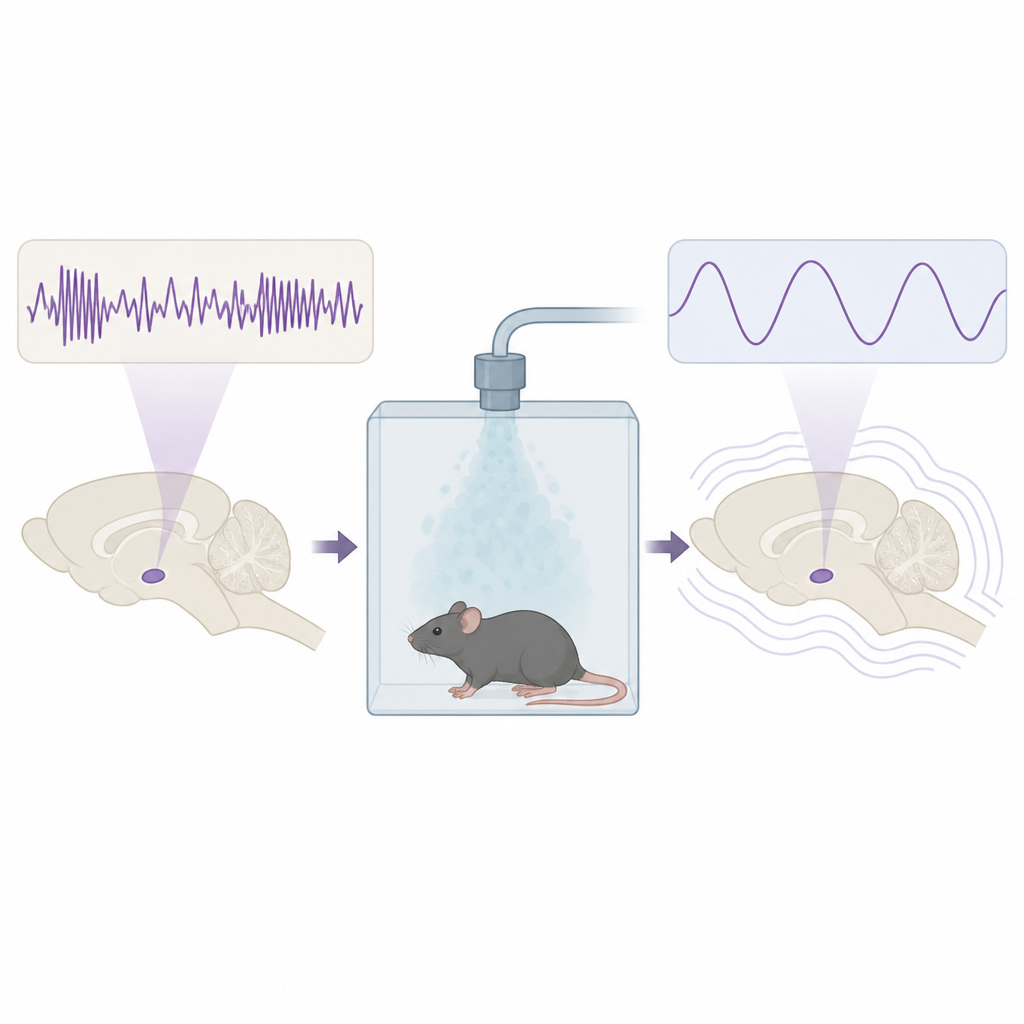
Mice were placed in a small chamber where they could move freely while breathing oxygen mixed with the anesthetic gas sevoflurane. The researchers recorded several types of signals at once: single cell spikes in the lateral hypothalamus, slow local waves around those cells, and surface brain waves from the cortex, along with muscle activity. The behavior of the animals was tracked using a simple test of whether they could right themselves if tipped over. This created a clear timeline of three stages: awake baseline, loss of the righting reflex under anesthesia, and recovery of that reflex as they emerged.
Different neuron groups respond in different ways
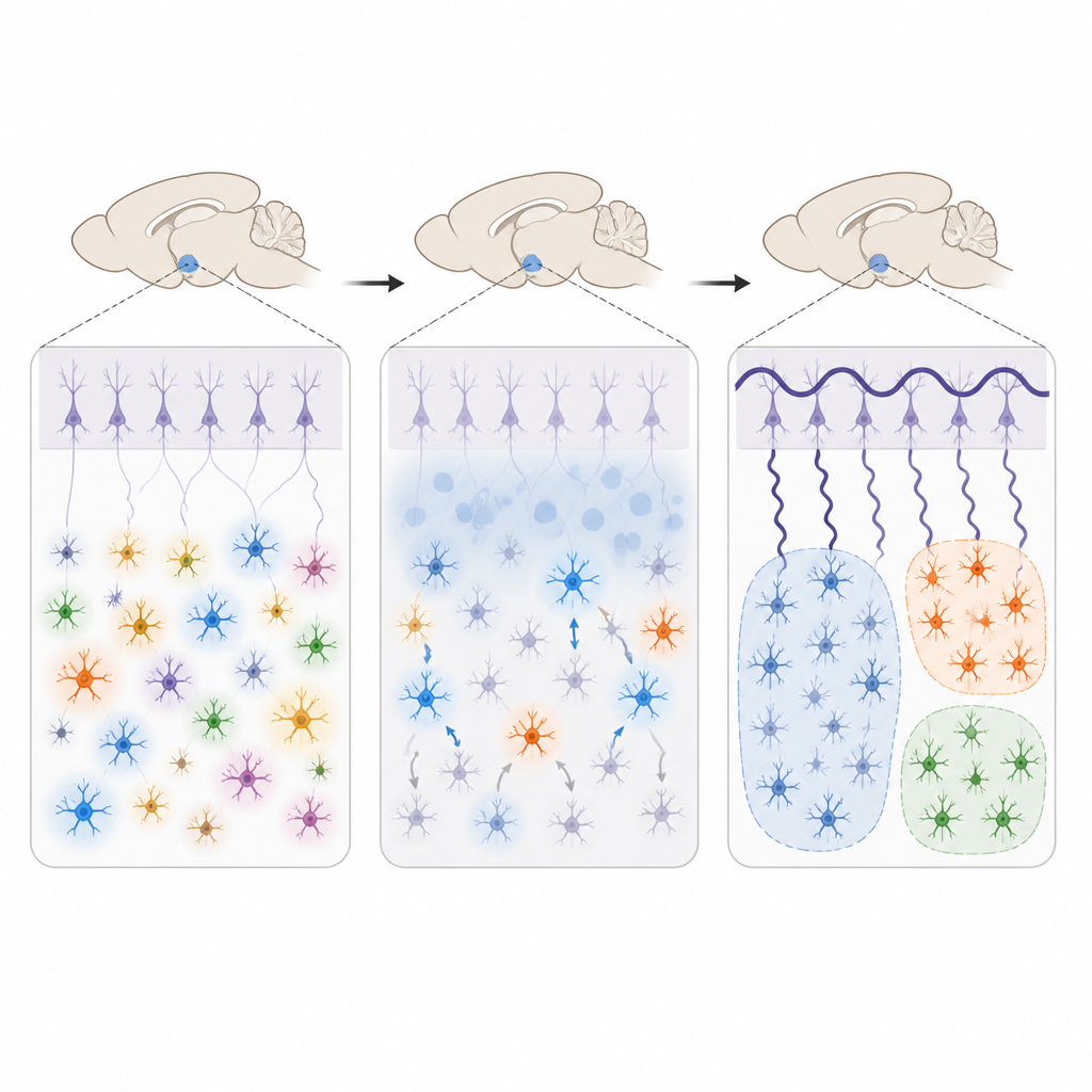
Zooming in on individual cells, the team found that most lateral hypothalamus neurons fell into one of three response patterns when sevoflurane took effect. About four out of five cells sharply reduced their firing during anesthesia and recovered as the animals woke up. A smaller group showed the opposite pattern, becoming more active under the drug, while a third group stayed largely unchanged. The suppressed cells tended to have larger and more distinctive electrical spikes in the awake state, suggesting they may belong to a particular functional class. In some cells, both the size of each spike and the firing rate changed together across states, hinting that anesthesia alters not only how often neurons fire but also the shape of their electrical signals.
Slow waves and tighter brain-wide coordination
At the level of larger networks, the study showed that sevoflurane shifted brain activity toward slow, high-amplitude waves in both the cortex and the lateral hypothalamus, similar to deep sleep. In the hypothalamus, very low-frequency power was especially strong, pointing to highly synchronized local activity. The cortex, by contrast, showed stronger relative shifts across a broader range of frequencies, indicating that it is more sensitive overall to anesthetic depth. Importantly, the slow rhythms in the hypothalamus and cortex became more tightly linked during anesthesia, as if these distant regions were marching in lockstep. This extra coordination faded again as the animals woke up.
What these findings mean for understanding anesthesia
Put in simple terms, the work paints a multi-layered picture of how a key wakefulness hub in the brain quiets down and changes rhythm under sevoflurane, while also becoming more tightly coupled to slow, sleep-like waves across the cortex. Rather than acting as a simple on-off switch, the lateral hypothalamus appears to reorganize its cell groups and join a broader slow-wave network during anesthesia. These detailed measurements of single cells and brain waves together may help explain why anesthetic drugs resemble deep sleep in some ways and could guide future efforts to monitor and control how patients enter and leave unconscious states.
Citation: Li, Q., Jia, Q., Song, Y. et al. State-dependent neuronal and network dynamics in the lateral hypothalamus across sevoflurane anesthesia–emergence revealed by microelectrode arrays. Microsyst Nanoeng 12, 195 (2026). https://doi.org/10.1038/s41378-026-01283-4
Keywords: anesthesia, lateral hypothalamus, brain waves, neuronal firing, sevoflurane