Clear Sky Science · en
Detection of mitochondrial bioenergetics using a novel bimodal 3D microelectrode array (MEA)-based biosensor
Why tiny power plants matter
Every cell in your body relies on mitochondria, tiny structures often called the cell’s power plants, to keep its energy flowing. When these power plants falter, the result can be a wide range of illnesses, from heart disease and diabetes to cancer and neurodegeneration. This study introduces a new kind of miniature sensor that can “listen in” on mitochondria without using dyes or labels, offering a fresh window into how they work and how they fail as we age or develop disease.
A new way to listen to cell power
The researchers built a small, chip based device that combines two kinds of electrical measurements in one platform. At its heart is a three by three grid of needle like microelectrodes that stick up from the chip surface, forming a three dimensional microelectrode array. Instead of recording from flat cell layers, these tiny towers reach up into a compact mound, or pellet, of isolated mitochondria. The same electrodes can perform both passive impedance readings, which sense how easily electricity moves through the sample, and active voltage recordings, which pick up fast electrical events across mitochondrial membranes. 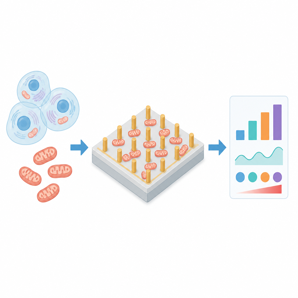
Building the tiny towers
To create this sensor, the team used digital light 3D printing to form plastic chips with tall pillars and matching holes for spring loaded connectors. They then coated the chip with thin layers of metals such as titanium, gold, and silver to make the pillars and surface conductive, and used laser machining to carve out separate electrode areas. A plastic insulation layer and a small culture well were added so that mitochondrial samples could be placed securely over the electrodes. By carefully tuning the height, spacing, and diameter of the pillars, the researchers produced arrays that not only fit within typical lab equipment but also reached deeply into the mitochondrial pellet, improving the quality and strength of recorded signals compared to flat, two dimensional electrodes.
Seeing and sensing live mitochondria
Before measuring electrical behavior, the team confirmed that their isolated mitochondria were alive and responsive. They stained the organelles with a fluorescent dye that glows more brightly when a voltage exists across the inner mitochondrial membrane, a hallmark of active energy production. As they supplied increasing amounts of fuel like succinate, glutamate, and malate, the fluorescent signal rose, signaling healthy, working mitochondria. The same fuel rich solutions were then used during electrical tests to mimic real cellular conditions and to see how mitochondrial activity changed the electrical properties that the chip could detect. 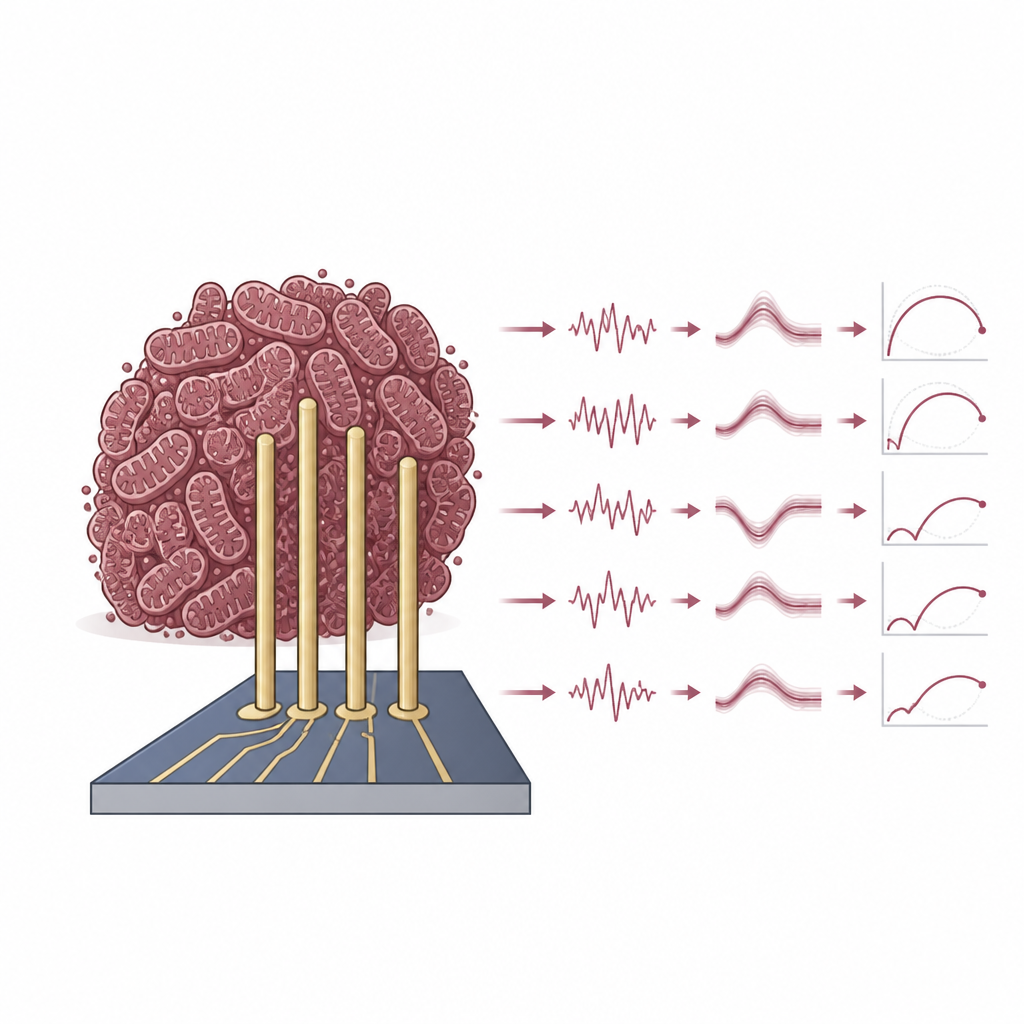
Tracking energy flow with impedance and voltage
Using electrochemical impedance spectroscopy, the researchers swept a gentle, alternating electrical signal across a wide range of frequencies and observed how the mitochondria responded. Adding a mitochondrial pellet to the buffer solution increased overall impedance, consistent with the insulating nature of their double membranes. When metabolic fuel was added, the impedance dropped slightly and the phase of the signal shifted, indicating that ion movement and membrane properties had changed as the electron transport chain turned on. Similar patterns appeared in mitochondria from both mouse fibroblast cells and human induced pluripotent stem cells, although their exact values differed. In separate experiments, time resolved voltage recordings from the mitochondrial pellet revealed tiny, rapid voltage oscillations that changed with fuel dose, suggesting real time activity in channels on the outer membrane or shifts in the inner membrane potential.
What this means for future health research
This work shows that a compact 3D microelectrode chip can measure how groups of mitochondria handle energy in two complementary ways at once. By reading both overall electrical resistance and fast voltage flickers, the biosensor offers a label free method to monitor the health of these cellular power plants in real time. With further refinement and pairing to organ on a chip systems, such devices could help scientists study how mitochondrial problems arise in complex diseases, test new drugs that target energy metabolism, and track how aging or stress reshapes the inner life of our cells.
Citation: James, R.K., Hostios, T.C., Chang, J. et al. Detection of mitochondrial bioenergetics using a novel bimodal 3D microelectrode array (MEA)-based biosensor. Microsyst Nanoeng 12, 208 (2026). https://doi.org/10.1038/s41378-026-01275-4
Keywords: mitochondria, biosensor, microelectrode array, bioenergetics, electrochemical impedance