Clear Sky Science · en
An advanced TMR sensor-based magnetrode for in vivo LFP magnetic field recording
Listening to the brain without touching its sparks
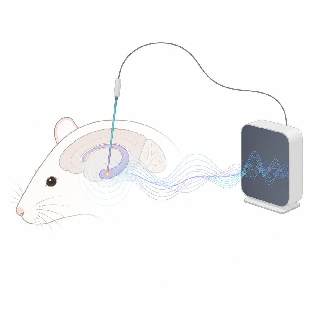
Our brains are buzzing with tiny electrical storms every second, and learning to read these storms could power future brain–computer interfaces that help people move, communicate, or even play games using thought alone. This study introduces a new way to listen in on brain activity, not by measuring electricity directly, but by sensing the faint magnetic fields that brain cells create, using a hair–thin device called a magnetrode.
A new kind of tiny brain probe
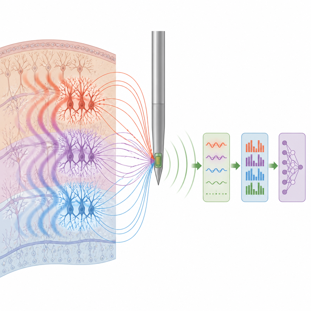
The researchers built a miniature probe based on tunneling magnetoresistance, a technology originally developed for advanced magnetic sensors. The active tip of their device is only tens of micrometers across, small enough to be implanted into the brain with limited damage. Instead of recording voltage like a traditional electrode, this magnetrode reacts to tiny magnetic fields produced when groups of neurons in a nearby region fire together. These combined signals, called local field potentials, reflect how networks of brain cells coordinate during movement, memory, and disease. The team carefully shaped and interconnected the sensor elements to keep the probe sensitive while reducing unwanted magnetic lag, so it could follow slow and fast changes in brain activity.
Seeing the faintest brain signals
Because the magnetic fields from neurons are extremely weak, the sensor must be quiet enough to pick them out from electronic noise. The authors measured how much random fluctuation the device produced at different frequencies and electrical drive settings. They found that low–frequency "1 over f" noise dominated the range where many brain rhythms live. By lowering the bias current that powers the device and switching from steady to high–frequency alternating drive, they showed that this troublesome noise can be strongly suppressed. The resulting detection limits, only a few nanotesla at one cycle per second and even smaller at higher frequencies, compared favorably with earlier implantable magnetic probes and with far bulkier magnetic–field instruments that cannot be implanted.
Testing with artificial and real brain signals
To check whether their probe could faithfully track local field potentials, the team first created a controlled test in the lab. A thin copper wire, driven by a specialized neural signal generator, mimicked the coordinated currents of a small group of neurons. The magnetrode sat close to this wire inside a shielded container, and its output was amplified, filtered, and then mathematically reconstructed. After processing, the magnetic signal closely matched the reference local field potential pattern, showing that the sensor and its electronics could recover the shape and timing of these slow brain rhythms.
Listening inside a living brain
The most important test came in living rats. The researchers gently implanted the magnetic probe and a standard microelectrode less than one–tenth of a millimeter apart in the hippocampus, a deep brain region involved in memory. Because both devices sampled nearly the same cluster of neurons, the electrical and magnetic recordings could be directly compared. Over several 100–second segments, the team analyzed the strength of different frequency bands in both signals. The magnetic and electrical spectra rose and fell together across the key brain rhythms, especially in the theta and beta ranges, and a statistical measure of similarity remained high and consistent. By contrast, recordings made from the magnetrode before implantation, when it only picked up background noise, showed much poorer agreement with the electrical signals, confirming that the in–brain magnetic traces truly reflected neural activity.
Built to survive the brain environment
Any implant must remain stable in warm, salty brain fluid. To test durability, the magnetrodes were soaked in artificial cerebrospinal fluid at body temperature for a week. The team repeatedly measured how strongly the device responded to test magnetic fields and how much its resistance changed. Both sensitivity and signal strength drifted by less than a few percent, suggesting that the protective layers around the sensor effectively blocked corrosion and that the probe could deliver reliable readings over the time scales needed for typical experiments.
What this means for future brain interfaces
This work shows that a tiny implanted magnetic sensor can track the same brain rhythms that standard electrodes see, while taking advantage of how magnetic fields pass more cleanly through tissue. For lay readers, the key idea is that the brain’s activity can be monitored not only by touching its electrical charges, but also by feeling their magnetic echoes. The magnetrode developed here is compact, sensitive, and stable enough to be used as a new kind of listening device for the brain, potentially enriching tools for brain–computer interfaces and for studying disorders linked to abnormal neural rhythms.
Citation: Wang, Y., Luo, J., Zhang, C. et al. An advanced TMR sensor-based magnetrode for in vivo LFP magnetic field recording. Microsyst Nanoeng 12, 177 (2026). https://doi.org/10.1038/s41378-026-01262-9
Keywords: brain magnetic recording, local field potentials, tunneling magnetoresistance, neural interfaces, brain computer interface