Clear Sky Science · en
Multispectral extended depth-of-field fluorescence microscopy with co-designed meta-optics and neural reconstruction
Seeing More in a Single Snapshot
Modern biology often relies on fluorescence microscopes to watch living cells at work, but there is a catch: only a very thin slice of a thick sample looks sharp at any one time. This study introduces a new approach, called MANTIS, that lets scientists capture crisp color images through the full thickness of tiny tissues in a single shot, reducing both wait time and light damage to living samples. 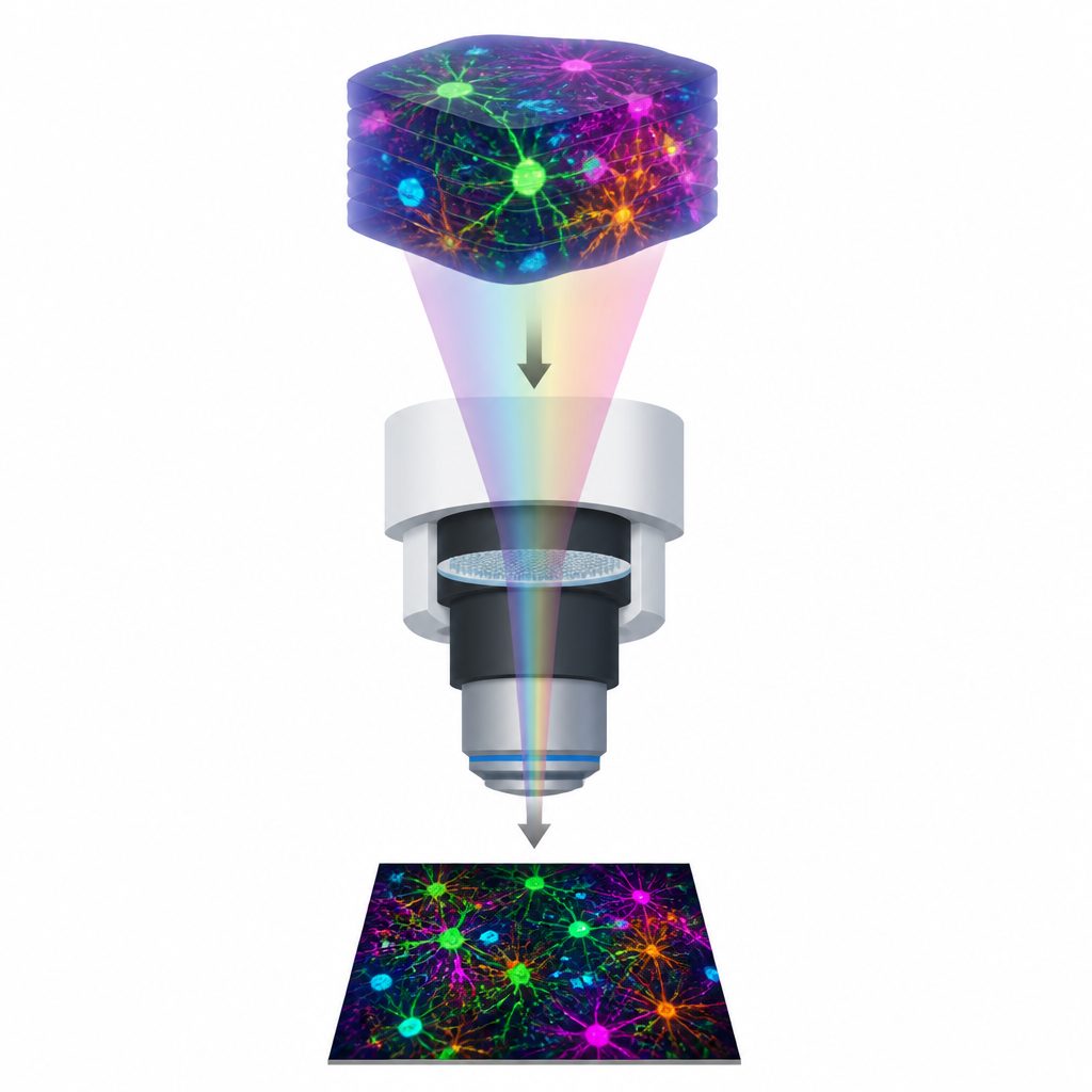
Why Thick Samples Are Hard to Image
Fluorescence microscopes highlight specific parts of cells using glowing dyes, revealing structures such as DNA, skeleton-like fibers, and membranes. To see very fine details, these microscopes use lenses that gather a lot of light, but this also shrinks the depth of field, the region that appears sharp. In thick samples, like tissue slices or 3D cell clusters, only a narrow layer is in focus, while structures above and below turn into blur. Traditional solutions move the sample or lens up and down, taking many pictures at different depths and then combining them, which is slow, creates motion artifacts, and exposes cells to repeated bursts of light.
Color Adds Another Layer of Trouble
Many biological experiments rely on several fluorescent colors at once, each marking a different molecule or structure. However, light of different colors does not pass through lenses in exactly the same way, so their best focus planes do not perfectly overlap. In thick samples this leads to some colors being sharp while others are smeared at the same depth, making it harder to tell whether two markers truly sit in the same place. Existing tricks to extend depth of field or split light into multiple views often sacrifice resolution, require complex hardware, or still depend on scanning through depths or colors.
A Thin Patterned Lens and a Smart Algorithm
The MANTIS system tackles these limits by jointly designing the optics and the image reconstruction. The microscope includes an ultrathin patterned surface made of tiny pillars placed at a key plane in the light path. This “meta-optic” reshapes how light from different depths and colors spreads on the camera sensor, making the recorded blur carry useful information across a much thicker region than usual. A physics-guided neural network then learns how to decode these blur patterns back into sharp images for all wavelengths at once. The researchers train both the meta-optic design and the neural network together on realistic data, so the two stages become tuned partners rather than separate components. 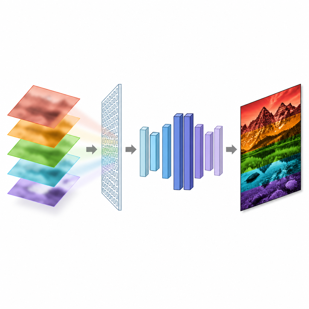
From Simulations to Real Cells and Tissues
Using simulations, the team explored how far they could extend the depth of field without losing too much detail, targeting ranges up to 75 micrometers with a very strong focusing lens. They showed that MANTIS could keep image quality relatively stable across depth and across four distinct color channels, accepting small trade offs in sharpness as the depth range grows. They then fabricated the meta-optic and installed it into a modified fluorescence microscope. Tests with glowing beads confirmed that the blur changed less with depth and color than in a standard system. When imaging flat cell layers, 3D cell spheroids around 50 micrometers thick, and mouse kidney tissue, the MANTIS reconstructions preserved cell outlines, nuclei, and fine structures across the sample thickness in a single exposure, while conventional images quickly became hazy away from the focal plane.
What This Means for Future Microscopy
In everyday terms, MANTIS lets researchers get a clear, multispectral view through a small 3D biological sample without having to refocus and stack many frames. By using a custom patterned surface together with a trained neural network, the system balances depth, detail, and color consistency in ways that conventional lenses alone cannot. While it does not separate individual depth layers like some scanning methods, it offers a practical route to fast, high resolution, all in focus fluorescence imaging, and can be adapted in the future to broader color ranges, thicker samples, and full 3D reconstruction.
Citation: Atalay Appak, I.A., Singh, H.J., Korpela, S. et al. Multispectral extended depth-of-field fluorescence microscopy with co-designed meta-optics and neural reconstruction. Light Sci Appl 15, 242 (2026). https://doi.org/10.1038/s41377-026-02337-y
Keywords: fluorescence microscopy, extended depth of field, meta-optics, computational imaging, multispectral imaging