Clear Sky Science · en
Ultra-wide-field, deep, adaptive two-photon microscopy for multi-scale neuronal imaging
Seeing the Brain in a New Way
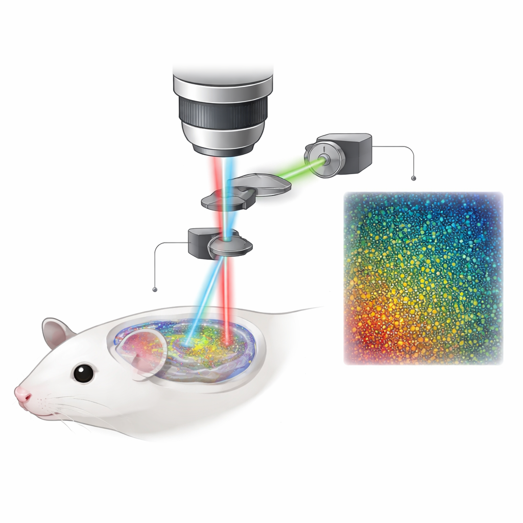
Understanding how the brain works means watching huge numbers of nerve cells communicating across many regions at once, not just zooming in on a tiny patch. Until now, brain microscopes have forced scientists to choose between seeing a wide area or peering deep below the surface with fine detail. This paper introduces a new microscope, called ULTRA, that largely breaks that trade-off, letting researchers watch tens of thousands of neurons firing across much of a mouse’s cortex, and down into deeper layers, all in a single experiment.
Why Regular Brain Microscopes Fall Short
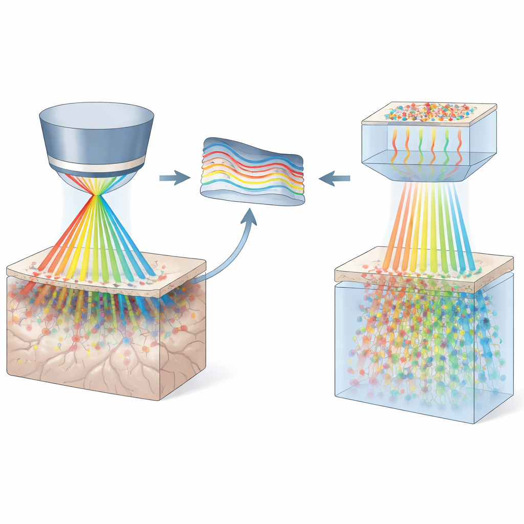
Two-photon microscopes are the workhorses of modern brain imaging because they can see activity deep inside living tissue with sharp detail. However, most can only view about a millimeter across at once, and their performance degrades as they try to look deeper or wider. Basic rules of optics say that if you make the field of view very wide, you usually have to give up on either resolution or light-gathering power. On top of that, the glass lenses must handle a wide range of colors without blurring, and every added lens piece can dim and distort the image. As a result, many existing wide-field systems are complex, expensive, and still struggle to combine very large area, depth, and single-cell clarity.
A New Microscope Built for Scale
The ULTRA system tackles these limits with a holistic redesign of the optical path. The authors created a custom water-immersion objective that can view an 8-millimeter-wide circle of brain surface—more than 50 square millimeters—while still resolving individual cells and reaching nearly a millimeter deep. Instead of treating the objective and downstream lenses separately, they optimized them together to keep blur and color errors very small across the entire field. Special glass types and carefully arranged lens groups help flatten the image and keep focus sharp from center to edge. A custom headplate holder lets the mouse’s skull be aligned so that many cortical areas sit at nearly the same depth, making it practical to scan wide areas in one go.
Sharper Views with Smart Optics and Better Detectors
ULTRA goes beyond clever lenses. It adds an adaptive mirror that can subtly reshape the incoming laser wavefront in real time to cancel distortions that grow worse toward the edges of a very wide field. This correction lets the system resolve tiny structures like dendritic spines several millimeters away from the center. On the detection side, the team built in large-area, high-output photomultiplier tubes that collect many more of the faint light particles returning from deep tissue. These detectors can handle strong and weak signals without saturating, improving the signal-to-noise ratio especially at depth and enabling fast scanning without sacrificing data quality.
Watching Large Brain Networks in Action
To show what ULTRA can do in real animals, the researchers imaged a variety of brain structures and activities in mice. They captured crisp pictures of inhibitory neurons, fine dendrites, and spines across a wide cranial window, and followed the same tiny spine for days, demonstrating stability. They also mapped blood vessels in four distant cortical regions at once and measured vessel diameters, lengths, spacing, and density. Most strikingly, they recorded calcium signals—a readout of neuronal activity—from many thousands of neurons spread over distances of 6–7 millimeters, both near the surface and in deeper layers around half a millimeter down. During locomotion, they could see how connections between brain regions strengthened and spread, revealing how movement changes large-scale cortical networks in real time.
What This Means for Brain Research
For a non-specialist, the key message is that ULTRA turns what used to be a narrow keyhole view of the brain into something closer to a panoramic window, without losing the ability to see single cells and fine branches. It can image a huge patch of the cortex, reach into deeper tissue, and follow activity changes during behavior, all with high clarity. Although there is still room to improve aspects like the vertical resolution and working distance for larger animals, this system already surpasses previous wide-field designs in the area and volume it can probe in living brains. ULTRA is likely to accelerate studies that link local cell behavior to brain-wide networks, bringing us closer to understanding how distributed groups of neurons work together to produce perception, movement, and memory.
Citation: Yang, M., Zhou, ZQ., Lang, S. et al. Ultra-wide-field, deep, adaptive two-photon microscopy for multi-scale neuronal imaging. Light Sci Appl 15, 198 (2026). https://doi.org/10.1038/s41377-026-02252-2
Keywords: two-photon microscopy, neural imaging, cortical networks, adaptive optics, brain-wide activity