Clear Sky Science · tr
Kesilmiş Yaratıcı Kurgu: Kedi Balığı Üzerinde Çalışmalar
Why young brains and head injuries matter
Head injuries are a leading reason children end up in the emergency room, especially those under five years old. Even when scans look normal, many of these children go on to have problems with memory, balance, and attention because the wiring inside the brain has been stretched or torn. To understand what actually happens inside a young, developing brain after a bump to the head—and how this might shape problems later in life—researchers need animal models that closely mimic a child’s brain rather than an adult’s.
A small animal that thinks big
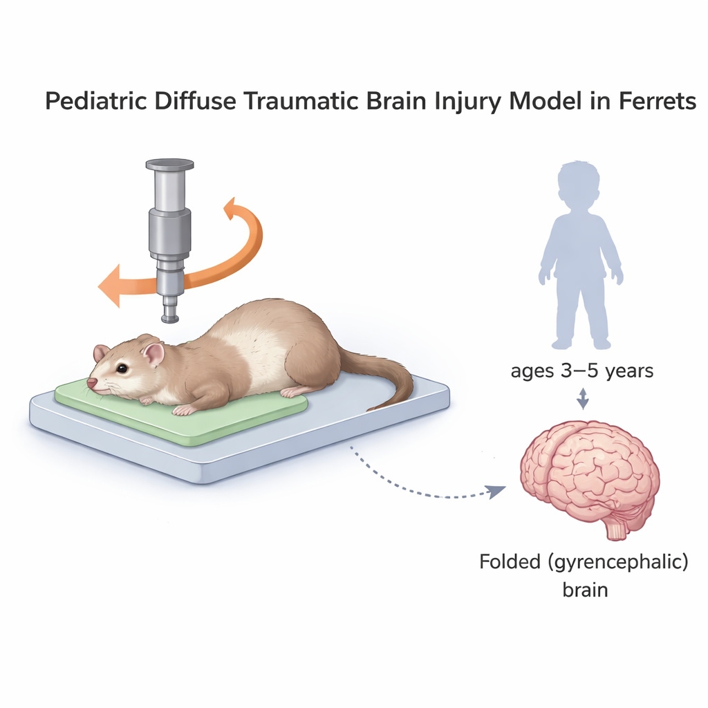
Most laboratory studies of brain injury use rats and mice. Their brains are smooth and have relatively little white matter, the “cables” that connect different brain regions. Human brains, in contrast, are heavily folded and rich in white matter. Ferrets, like humans, have folded brains with substantial white matter, but they are much smaller and easier to house than pigs, the other common large-brain model. In this study, scientists worked with 2–3‑month‑old ferrets whose brain development roughly matches that of 3–5‑year‑old children. They adapted a device called CHIMERA, which delivers a controlled strike to the skull and causes the head to move and rotate—more like a real‑world fall or collision than a simple poke to one spot on the brain.
What happens inside the young brain’s wiring
The researchers examined the ferrets’ brains up to 72 hours after injury. Instead of bruises or bleeding that you could see with the naked eye, the main damage was hidden inside the long, thin nerve fibers that carry signals between brain regions. Using special stains, they tracked two early warning signs in these fibers: a traffic jam of a normally moving protein (APP), and damage to structural “scaffolding” proteins (NFL) that help axons keep their shape. Within a day, there was a surge of APP build‑up in central white matter pathways such as the corpus callosum and fornix—major communication highways that support movement and memory. By three days, this particular signal had faded in many regions, but NFL‑related damage remained widespread, showing that some axons were still structurally compromised even after the initial traffic jams eased.
The brain’s immune response and blood clues
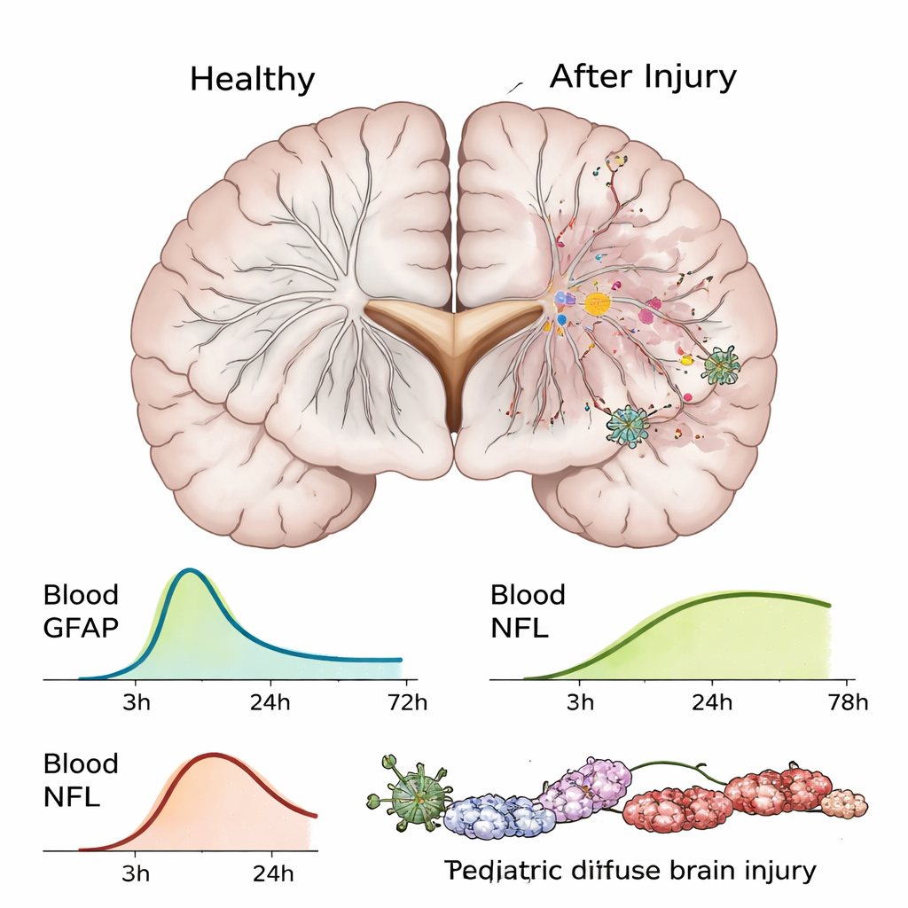
Beyond the nerve fibers themselves, the team looked at microglia, the brain’s resident immune cells. These cells changed shape and increased in number by 72 hours after injury, especially in the same white matter tracts that showed the most axonal damage and in deep regions such as the hypothalamus. This suggests that an inflammatory response builds up over days and could influence how the young brain recovers—or fails to recover—after trauma. The scientists also measured two proteins in the blood that are already being tested in injured children. GFAP, a marker of support cells in the brain, spiked within 30 minutes and stayed high for about a day before returning to normal by 72 hours. NFL, which reflects damage to long nerve fibers, was low in uninjured animals but rose sharply by 24 hours and remained elevated at 72 hours. These blood changes mirror patterns seen in pediatric patients and could help doctors time when and how to test for hidden brain damage.
Subtle problems with movement and memory
To find out what these microscopic changes mean in everyday terms, the ferrets were put through a series of simple tasks. In an open arena, their overall activity was similar to uninjured animals, suggesting they could still walk and explore. But on a narrow ladder, injured ferrets moved more slowly, hinting at balance and coordination problems. In puzzle‑based tasks that required learning, remembering, and adjusting to new rules, injured ferrets struggled more than their uninjured peers, especially when tasks became slightly harder. They were slower to remember where a reward had been and less flexible at adapting when the reward moved. These subtle difficulties resemble the balance and thinking problems often seen in young children after concussion, even when brain scans appear normal.
What this means for children with head injuries
This new ferret model shows that a blow to a young, folded brain can cause widespread damage to nerve fibers and spark an immune response, without obvious bruising or swelling. It reproduces key features of pediatric head injury: hidden white‑matter damage, short‑term spikes in blood markers, and mild but meaningful problems with movement and thinking. For families and clinicians, the work underscores that a “mild” head injury in a preschooler can still disrupt developing brain circuits in ways that may not show up on routine scans. For scientists, the model offers a practical way to test how early brain injuries evolve over time and to explore treatments that might protect or repair the brain’s wiring during a crucial window of development.
Atıf: Krieg, J.L., Hooper, C., Kapuwelle, H. et al. Development of a paediatric model of diffuse traumatic brain injury in ferrets. Sci Rep 16, 6037 (2026). https://doi.org/10.1038/s41598-026-37303-6
Anahtar kelimeler: pediatric traumatic brain injury, diffuse axonal injury, ferret brain model, white matter damage, brain biomarkers