Clear Sky Science · tr
Ritmik ve desenli TMS protokollerine karşı farklı nöral yanıtlar: EEG spektral analizinden çıkarımlar
Why brain stimulation patterns matter
Many people with major depression do not get enough relief from medications alone. Repetitive Transcranial Magnetic Stimulation (rTMS) offers new hope by using magnetic pulses at the scalp to nudge brain activity in helpful directions. But rTMS can be delivered in different “rhythms,” and patients often respond well to one style but not another. This study asked a simple but important question: do different rTMS pulse patterns actually drive the brain in different ways, and could that help explain why some people improve while others do not?
Two ways to tap the brain
The researchers focused on two common ways of giving rTMS to the front of the brain, over an area called the left dorsolateral prefrontal cortex, a key hub in mood and thinking. One method used steady, drum-like pulses at a single pace, called rhythmic stimulation. The other used brief bursts of very fast pulses grouped into patterns, called patterned stimulation. Sixteen adults with hard-to-treat depression received dozens of short trains of both types in a single session, covering a wide span of pulse speeds. During this “interrogation” session, the team recorded electrical activity from across the scalp using a 64-channel EEG cap to see how each pattern and pulse speed changed brain rhythms and the way regions talked to one another.
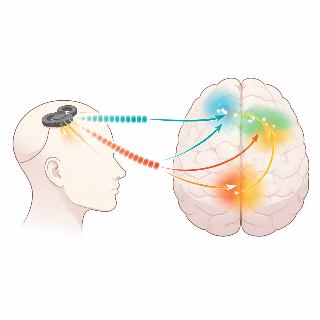
Listening to brain rhythms
Brain cells naturally fire in repeating waves, or oscillations, at different speeds that are linked to states such as drowsiness, focused attention, or emotional processing. The team split these rhythms into four bands from very slow to faster (delta, theta, alpha, and beta). For every short train of rTMS, they compared EEG signals in the second before and the second after stimulation. Using advanced mathematical tools, they estimated where in the brain these signals arose and how strongly the targeted prefrontal area was influencing about 100 other regions. They then used statistical models that could separate broad effects of stimulation from differences between individual patients.
Shared shifts and clear contrasts
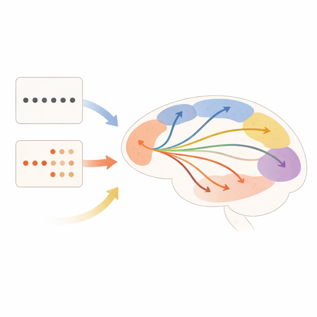
Both rhythmic and patterned pulses had widespread effects, even though they all hit the same spot on the scalp. Across most conditions, very slow (delta) and slow (theta) rhythms decreased after stimulation, while faster beta rhythms often rose, especially for patterned trains. However, the two styles were far from identical. Patterned stimulation produced the strongest boosts in beta power and drove changes in specific “inner” areas such as parts of the cingulate cortex and the precuneus, regions involved in self-focus and internal mentation. Rhythmic stimulation, especially at higher speeds, more strongly affected activity near the stimulation site and across broader swaths of cortex. In several cases, the exact prefrontal subregions and deeper midline structures that responded differed clearly between the two patterns.
Rewiring communication lines
Beyond local power changes, the study examined how stimulation altered communication pathways. Both styles strengthened the influence of the targeted prefrontal area on the orbitofrontal cortex and a midline region linked to mood regulation, suggesting a shared route through which rTMS might ease depressive symptoms. Yet only rhythmic pulses increased connectivity within nearby frontal regions, while only patterned trains reduced connectivity to the left precuneus and certain visual areas. As the pace of rhythmic pulses increased from slower to faster, connectivity changes spread more widely, particularly in higher-frequency response bands. In short, by tweaking how fast and in what pattern the pulses were delivered, the researchers could steer the same prefrontal “steering wheel” to different sets of downstream regions.
Toward more tailored brain stimulation
To a lay observer, all rTMS sessions may look the same: a magnetic coil clicking over the forehead. This work shows that under the surface, different pulse patterns and speeds can engage partly distinct brain circuits, even when aimed at the very same location. Both rhythmic and patterned approaches appear capable of strengthening helpful fronto-limbic connections, but they also sculpt separate pathways through networks involved in self-reflection, attention, and emotion. These mechanistic differences may help explain why one protocol works for some patients and not others. In the future, short “interrogation” sessions like the one used here could map how an individual’s brain responds to various rTMS settings, allowing clinicians to choose the pattern and frequency that best correct that person’s network imbalances and, ultimately, improve their chances of recovery.
Atıf: Valles, T.E., Shamas, M., Hawkins, H. et al. Differential neural responses to rhythmic and patterned TMS protocols: Insights from EEG spectral analysis. Neuropsychopharmacol. 51, 813–821 (2026). https://doi.org/10.1038/s41386-025-02306-w
Anahtar kelimeler: transkraniyal manyetik stimülasyon, majör depresif bozukluk, beyin ağları, EEG osilasyonları, nöromodülasyon