Clear Sky Science · en
Vibrational photothermal imaging: theory, instrumentation, and applications
Seeing Molecules Through Their Heat
Many breakthroughs in medicine and materials science depend on being able to see what molecules are doing inside cells, tissues, and tiny devices—ideally without adding any labels or dyes that might disturb them. This article reviews a rapidly evolving approach called vibrational photothermal imaging, which detects the faint bursts of heat that molecules release after absorbing light. By turning those tiny temperature changes into images, researchers can map chemistry inside living cells, batteries, plastics, and even historical paintings with remarkable sensitivity and fine detail.
From Light Absorption to Tiny Heat Bursts
When a molecule absorbs light, most of that energy does not come back out as glow; instead, it quickly turns into heat as the molecule relaxes. Vibrational photothermal imaging takes advantage of this universal effect. A carefully tuned infrared “pump” beam excites specific chemical bonds, and a second “probe” beam senses the resulting temperature rise as changes in how light passes through or scatters from the sample. Because vibrational excitations convert essentially all their energy to heat, this method is naturally sensitive and works without fluorescent tags. The authors explain how the temperature rises and falls over billionths to millionths of a second, and how heat slowly spreads through the surrounding medium, setting basic limits on speed and sharpness. 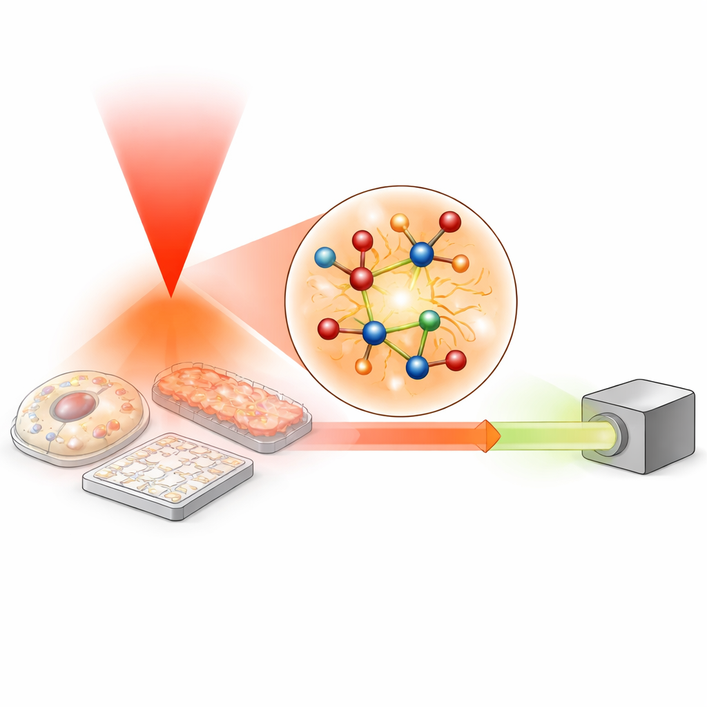
Turning Heat into Contrast
The review describes several clever ways to turn those tiny temperature changes into visible contrast. In some setups, the warmed region acts like a fleeting lens that slightly focuses or defocuses the probe beam. In others, the heat alters how much a particle scatters light, or shifts the optical phase—the precise “timing” of the light wave. Still others rely on fluorescent dyes whose brightness depends on temperature, or on sound waves generated when heated regions rapidly expand. Each mechanism offers its own trade-offs in sensitivity, resolution, and compatibility with living samples, but all rest on the same basic principle: local heating subtly changes optical properties, which can be read out as an image.
Building Microscopes Around Heat
To harness these effects, researchers have engineered a family of microscopes. In point-scanning instruments, tightly focused infrared and visible beams move across the sample to build up images with submicron resolution and fast spectral readout. Widefield systems instead illuminate larger areas and rely on cameras, using timing tricks to separate “hot” and “cold” frames so even nanosecond-scale heating can be captured with relatively slow sensors. Tomography schemes add multiple viewing angles and advanced computation to reconstruct three-dimensional chemical maps. The review also explains how the choice of light source, focusing geometry, and detection electronics must balance sensitivity, speed, and gentleness for living specimens.
Following Chemistry in Cells, Materials, and the Environment
Because photothermal signals are tied to specific molecular vibrations, these microscopes can distinguish many kinds of chemicals at once. The authors survey applications that range from tracking microbial metabolism and drug responses, to watching enzyme activity and lipid storage in individual cells, to mapping the structure of protein aggregates linked to neurodegenerative diseases. In tissues, the technique enables label-free “virtual staining” for pathology and high-resolution studies of bone, brain, and tumors. Beyond biology, it reveals nanoscale structure in perovskite solar cells, battery interfaces, catalysts, pharmaceuticals, and even pigments in masterpieces by van Gogh. Environmental scientists use it to identify micro- and nanoplastics, aerosols, and pollutants in water and soil, thanks to its ability to recognize polymers and contaminants down to hundreds of nanometers in complex mixtures. 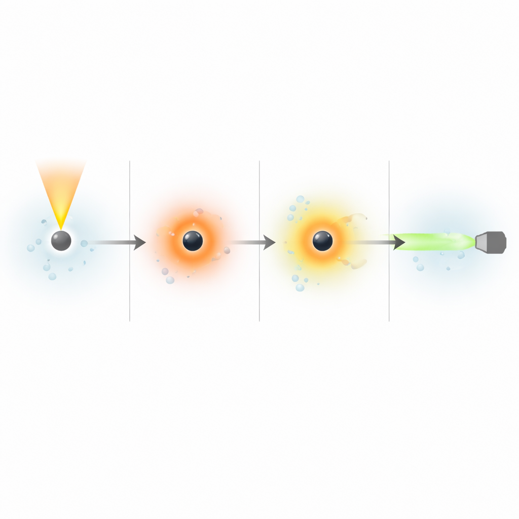
New Windows and Future Directions
The review also introduces newer variants that work at different wavelengths. Stimulated Raman photothermal microscopy uses near-infrared light to excite vibrations indirectly, producing stronger thermal signals while keeping optical noise low. Shortwave infrared photothermal imaging pushes deeper into tissue, achieving millimeter penetration while still resolving cell-scale structures. Looking ahead, the authors foresee faster imaging, higher resolution aided by computation and tailored beam shapes, and even extension to spectral regions such as X-rays and terahertz waves. They highlight prospects in clinical diagnosis—like rapid antimicrobial testing, improved cancer margins, and noninvasive metabolic monitoring—while emphasizing the need to manage heating to keep living systems safe. In essence, the field is learning to read chemistry by listening to heat, turning a universal side effect of light absorption into a powerful, label-free window on the molecular world.
Citation: Jiaze Yin, Pin-Tian Lyu, Rylie Bolarinho, Yifan Zhu, Xiaowei Ge, Hongli Ni, and Ji-Xin Cheng, "Vibrational photothermal imaging: theory, instrumentation, and applications," Optica 12, 1367-1387 (2025). https://doi.org/10.1364/OPTICA.564920
Keywords: vibrational photothermal microscopy, mid-infrared imaging, label-free chemical imaging, molecular spectroscopy, biophotonics