Clear Sky Science · en
Anisotropically multiplanar-focal photon-sieve splitter from extreme ultraviolet to soft X-ray
Looking at Tiny Worlds with New Light Tricks
Our modern world depends on technologies that can draw and inspect features far smaller than a speck of dust, from computer chips to advanced materials. To do this, scientists use very short-wavelength light, in a range called extreme ultraviolet and soft X-ray, which can reveal details far beyond what visible light can show. But shaping and splitting this kind of light is extremely hard, because most materials absorb it instead of bending or reflecting it cleanly. This paper introduces a new kind of ultra-thin optical device that can split and focus such light onto several spots at different depths, opening the door to sharper imaging and clever new measurement techniques.
A New Kind of Tiny Light Sieve
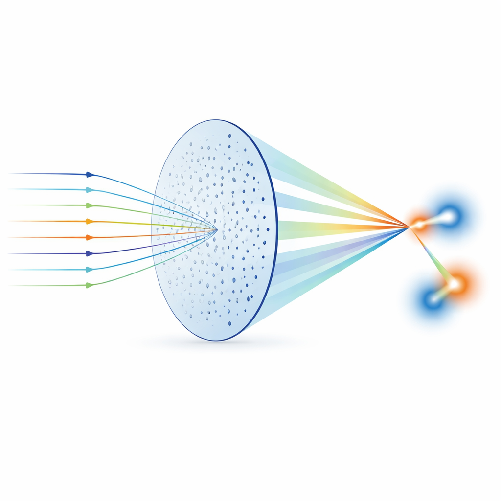
Instead of using traditional lenses or mirrors, the researchers rely on a concept called a photon sieve—a thin membrane drilled with thousands of carefully placed microscopic holes. When light passes through this pattern of holes, it is bent by diffraction and can be made to focus, somewhat like a lens but without the need for thick glass. Photon sieves are especially attractive for extreme ultraviolet and soft X-ray light, where normal optics fail because the materials soak up too much energy. By changing where the holes sit and how big they are, scientists can sculpt the light in intricate ways, making photon sieves a powerful alternative to conventional optics in this demanding wavelength range.
Splitting Light in Depth, Not Just Sideways
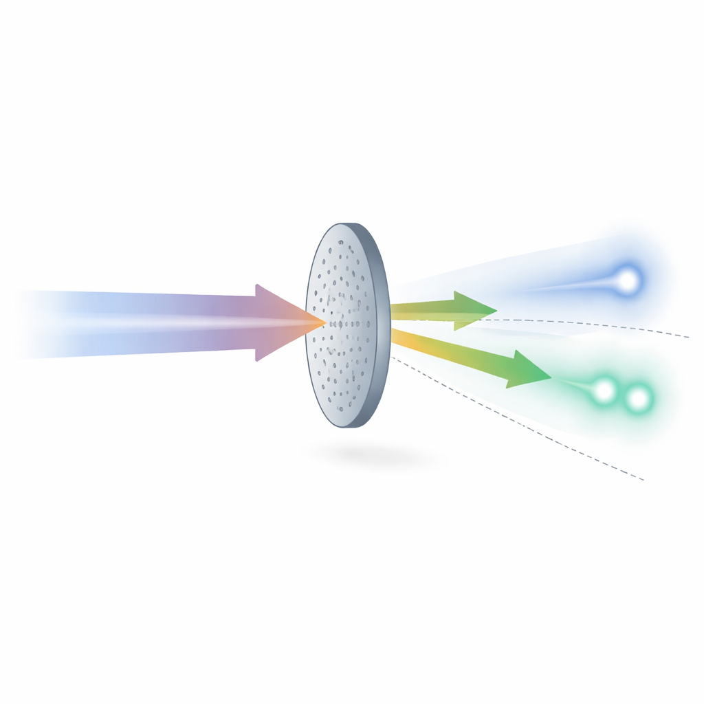
The main innovation of this work is a device the authors call an anisotropically multiplanar-focal photon-sieve splitter. In simpler terms, it is a photon sieve designed to create three separate bright spots of light that are not only apart from one another but also lie on two different focus planes along the beam path. One bright spot sits on a single-focus plane, while a pair of spots appears together on a second plane farther away. Achieving this requires encoding a special number pattern—based on an ancient “Greek ladder” sequence—into how the holes are arranged. The pattern is optimized using a computer algorithm that treats each possible layout as a “chromosome” and gradually improves it until the desired three-spot focusing behavior is reached.
Building and Testing the Ultra-Thin Splitter
To turn the design into reality, the team fabricated a photon-sieve splitter about 0.8 millimeters across on a very thin silicon nitride film, using microfabrication techniques similar to those used in chip making. Roughly half of the membrane is open holes, which keeps manufacturing relatively simple but also limits how efficiently it redirects the light. The splitter was then tested with a 46.9-nanometer extreme ultraviolet laser that delivers very short, intense pulses. A plastic material called PMMA served as a recording plate: the incoming light subtly alters its surface, and after processing, the surface shape directly reveals where the light was most intense. By mechanically scanning this plate along the beam direction and examining it with microscopes, the researchers could see how the focused spots changed size and position near each focus plane.
Checking That Focus Matches the Design
The raw images of the tiny craters and bumps in the PMMA showed that the three focal spots behaved as intended: as the recording plate moved through the beam, the spots shrank to a minimum size at a single-focus plane and at a second plane containing two spots. To measure this more precisely, the team used atomic force microscopy to map the surface in detail and then applied a numerical “autofocus” procedure. By digitally propagating the measured patterns back and forth in space using known diffraction formulas, they could find the distances where the spots became sharpest. The resulting spot sizes were just a few hundred billionths of a meter across and matched theoretical predictions closely, confirming that the splitter produced the correct focus positions and intensities despite small experimental imperfections.
Why This Matters for Future Imaging Tools
By showing that a single, flat, perforated membrane can reliably split extreme ultraviolet light into multiple focused spots at different depths, this work provides a new building block for advanced imaging and measurement systems. Such a splitter could let scientists capture several diffraction patterns in one shot, or compare slightly different focus planes without moving bulky optics, which is valuable for techniques like coherent diffraction imaging, phase diversity, and interferometry. In everyday terms, it is like having a paper-thin “light switchboard” that can send one powerful, hard-to-handle beam into several precise channels at once. This capability could help push the limits of how finely we can see and measure structures in the tiny worlds that underpin modern technology.
Citation: Keyang Cheng, Huaiyu Cui, Ziyi Zhang, Yuni Zheng, Dongdi Zhao, Qi Li, Yongpeng Zhao, and Junyong Zhang, "Anisotropically multiplanar-focal photon-sieve splitter from extreme ultraviolet to soft X-ray," Optica 12, 1388-1390 (2025). https://doi.org/10.1364/OPTICA.559913
Keywords: extreme ultraviolet optics, photon sieve, multifocal beam splitting, diffractive imaging, soft X-ray focusing