Clear Sky Science · en
Anterior segment optical coherence tomography (ASOCT) evaluation of cryo-preserved and vacuum dried amniotic membrane used in the management of persistent corneal epithelial defects (PED)
Why this matters for eye health
When a scratch or ulcer on the clear front window of the eye refuses to heal, people can suffer pain, blurred vision and even risk blindness. This study looks at two different ways of using a special biological “bandage” made from the amniotic membrane (part of the placenta) to help stubborn corneal wounds close. The researchers also use a high‑tech eye scan to watch the surface of the eye recover in fine detail.
A natural bandage for a stubborn eye wound
The transparent surface of the eye, the cornea, is covered by a thin skin of cells that normally repairs itself quickly after minor injuries. In some people, however, this outer layer breaks down or fails to grow back; this is called a persistent epithelial defect (PED). PEDs can arise after infections, nerve damage, surgery, or other eye diseases, and they raise the risk of scarring, infection and even holes in the eye. Doctors increasingly turn to amniotic membrane transplantation, in which a thin layer from the placenta is laid onto the damaged cornea to encourage healing and reduce inflammation.
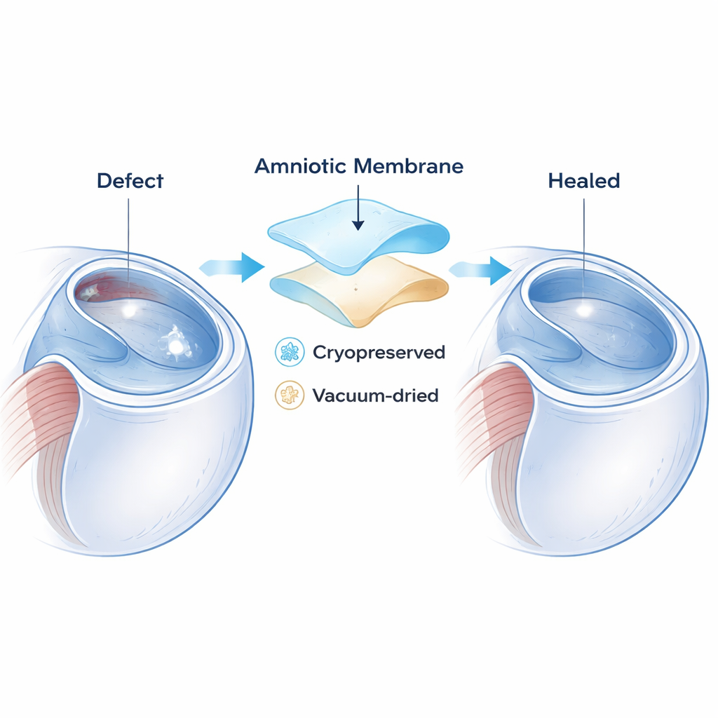
Two ways to prepare the same healing tissue
Amniotic membrane can be preserved in different ways before it is used. One method is cryopreservation, where the tissue is frozen at very low temperatures to keep its natural molecules as intact as possible. Another is to dry the tissue under vacuum after adding protective sugars; this makes a shelf‑stable product (known commercially as Omnigen) that can be stored at room temperature and used straight from the box. Both versions are thought to contain growth factors and other helpful substances, but until now no study had carefully compared how they perform in patients with persistent defects by actually measuring changes in the cornea over time.
Watching healing in real time with detailed eye scans
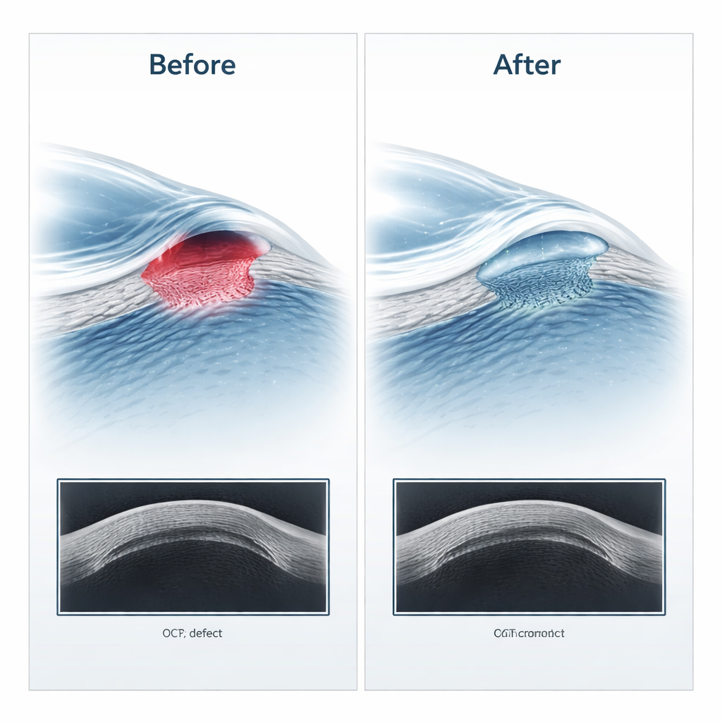
The team reviewed 29 patients treated at a UK eye hospital between 2017 and 2024. Fourteen eyes received cryopreserved membrane and 15 received the vacuum‑dried version. In all cases, surgeons placed several thin layers of the membrane into the ulcer so that it acted as a graft filling the defect, rather than just a temporary patch on top. Patients were followed with slit‑lamp examinations, special dye to highlight remaining defects, and anterior segment optical coherence tomography (ASOCT)—a non‑contact scan that slices the cornea into high‑resolution cross‑sections. Measurements were taken before surgery, about one week afterward, and again at around three to four weeks, once the surface had either healed or failed.
Similar healing, regardless of the membrane type
Just over three‑quarters of the eyes (22 out of 29) healed successfully. The rate of success was similar for both cryopreserved and vacuum‑dried membranes. In the eyes that healed, ASOCT showed that the underlying corneal bed became significantly less swollen and thick over time, reflecting resolution of inflammation and fluid, while the total thickness of the cornea—including the transplanted membrane—stayed relatively stable once healing was complete. The outermost cell layer regained a near‑normal thickness in both groups, and there were no meaningful differences in any measured thickness between the two membrane types at any stage. In other words, both preparations supported corneal repair to a similar degree.
What this means for patients and clinics
For patients facing a persistent corneal wound, this study suggests that doctors can choose either frozen or vacuum‑dried amniotic membrane without sacrificing healing potential. The dried product has practical advantages, such as easier storage and quicker availability, which may be important in busy hospitals or regions without ready access to tissue banks. The work also highlights how detailed eye imaging can give doctors objective, numerical information on how well the cornea and the graft are recovering, and could even support remote, image‑based follow‑up in the future. Overall, the message is reassuring: both forms of this natural eye bandage appear equally helpful in getting stubborn corneal defects to close and protecting vision.
Citation: ElZawahry, F.O., Rossi, C., Sahay, P. et al. Anterior segment optical coherence tomography (ASOCT) evaluation of cryo-preserved and vacuum dried amniotic membrane used in the management of persistent corneal epithelial defects (PED). Eye Open 2, 8 (2026). https://doi.org/10.1038/s44440-025-00007-3
Keywords: amniotic membrane, corneal ulcer, persistent epithelial defect, optical coherence tomography, ocular surface disease