Clear Sky Science · en
Substrate-assisted cathodoluminescence
A gentler way to see the tiniest lights
Modern electron microscopes can make materials glow, revealing how light behaves on the smallest scales. But the same high-energy electrons that create this glow can also damage delicate quantum emitters that might power future sensors and quantum technologies. This paper explores a subtler approach: using electrons that are first scattered by the supporting substrate to excite light emitters in diamond, allowing scientists to probe them with far less disturbance.
How electron microscopes make things shine
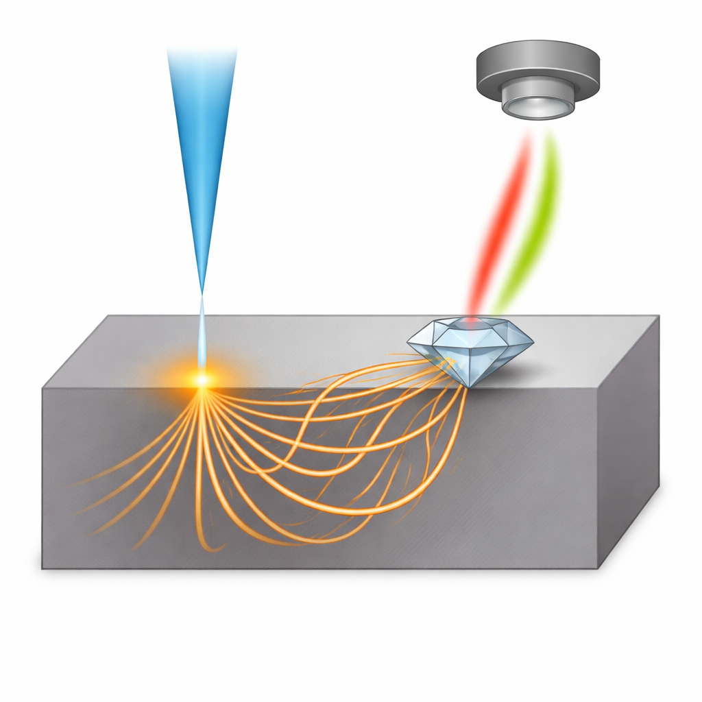
In cathodoluminescence microscopy, a focused beam of fast electrons hits a sample and makes it emit light. This technique is prized because it combines high spatial resolution with spectral and timing information, letting researchers study tiny light sources such as color centers in diamond. Traditionally, the electron beam either strikes the emitter directly, or passes very close by so that its electromagnetic field excites the material without actual impact. A third route has been hinted at but not well understood: indirect excitation, where electrons first interact with the underlying substrate and only then reach the emitter. The authors set out to clarify how this indirect pathway works and how far its influence extends.
Letting the substrate do the work
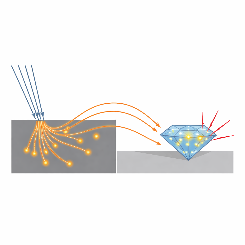
The team used microscopic diamond crystals containing silicon-vacancy centers—bright, stable defects that act as tiny light sources—as local probes. In one set of experiments, they placed the electron beam directly on a diamond crystal and recorded its light spectrum and photon statistics. In another, they moved the beam a few micrometers away, onto the neighboring metal surface, making sure the beam never touched the diamond itself. Surprisingly, the diamond still lit up with a spectrum very similar to the directly excited case, even though the light intensity dropped by about a factor of one hundred. At the same time, the statistics of the emitted photons changed dramatically: the photons arrived in stronger bursts, a signature that the effective excitation rate experienced by the emitters had become much lower.
Backscattered electrons as hidden messengers
To uncover the physical carriers of this indirect excitation, the authors systematically varied the substrate material and the energy of the electron beam. They compared thin silicon nitride membranes with much thicker silicon frames, and also tested substrates such as silicon, germanium, graphite, and gold, which differ in atomic weight and density. Spatial maps of the diamond’s glow revealed broad halos extending several micrometers from the beam position, whose shapes changed in predictable ways with material and energy. These patterns matched what is expected for backscattered electrons—high-energy electrons that bounce around inside the substrate and re-emerge near the surface—rather than for low-energy secondary electrons, which travel only nanometer-scale distances. In light substrates such as silicon or graphite, the glow spread out with a smooth, bell-shaped profile, while in heavier materials like germanium and gold it dropped off more sharply, consistent with backscattering theory.
Measuring an invisible current with photon timing
Because the instrument can only measure the incoming beam current, not the tiny fraction that actually reaches the emitters indirectly, the researchers turned to photon-correlation measurements. They analyzed how strongly the emitted photons bunched together in time—a quantity that is known to vary inversely with the rate of electron impacts on the emitters. By recording this photon bunching for different beam currents and for various beam-to-diamond distances, they could infer the “effective” current that the emitters felt under indirect excitation. The data showed that direct and indirect excitation follow the same basic mechanism, but in the indirect case the effective current drops by several orders of magnitude as the distance grows, reaching values below one-tenth of a picoampere.
Why this matters for fragile quantum materials
These findings reveal that the substrate in an electron microscope is not just a passive support, but an active partner that can deliver a faint, extended shower of electrons to nearby emitters. By choosing the right substrate material and beam energy, researchers can engineer how far and how strongly this indirect excitation reaches, effectively tuning a gentle illumination field around sensitive samples. The work shows that substrate-assisted cathodoluminescence can probe quantum emitters with much lower damage risk while preserving their intrinsic light-emission properties, opening a path to more careful, spatially controlled studies of nanoscale light sources in future quantum and nanophotonic devices.
Citation: Ebel, S., Mortensen, N.A. & Morozov, S. Substrate-assisted cathodoluminescence. npj Nanophoton. 3, 18 (2026). https://doi.org/10.1038/s44310-026-00116-6
Keywords: cathodoluminescence, electron microscopy, quantum emitters, diamond color centers, backscattered electrons