Clear Sky Science · en
Single capture quantitative oblique back-illumination microscopy
Seeing Living Cells Without Dyes
Modern medicine increasingly depends on watching living cells in action, but most microscopes still need fluorescent dyes or slow scanning methods that can disturb tissues. This study introduces a new way to take sharp, three-dimensional pictures of living tissue with a single camera snapshot and no labels, potentially letting doctors and researchers watch blood flow and cellular changes in real time, right inside the body.
A Faster Way to Look Inside Thick Tissues
Many powerful imaging tools face a trade-off: some scan fast but miss fine details, while others reveal rich cellular structure but are slow or limited to thin samples on glass slides. An earlier technique called quantitative oblique back-illumination microscopy (qOBM) solved part of this problem by sending light into tissue from above, letting scattered light act like a hidden light source inside thick, cloudy samples. qOBM can measure how much the light wave is delayed by cells—a property linked to their internal structure—across three dimensions. But traditional qOBM needed four separate camera exposures from different lighting angles, which slowed it down and made it vulnerable to blurring whenever the sample moved.
Teaching a Microscope to Think
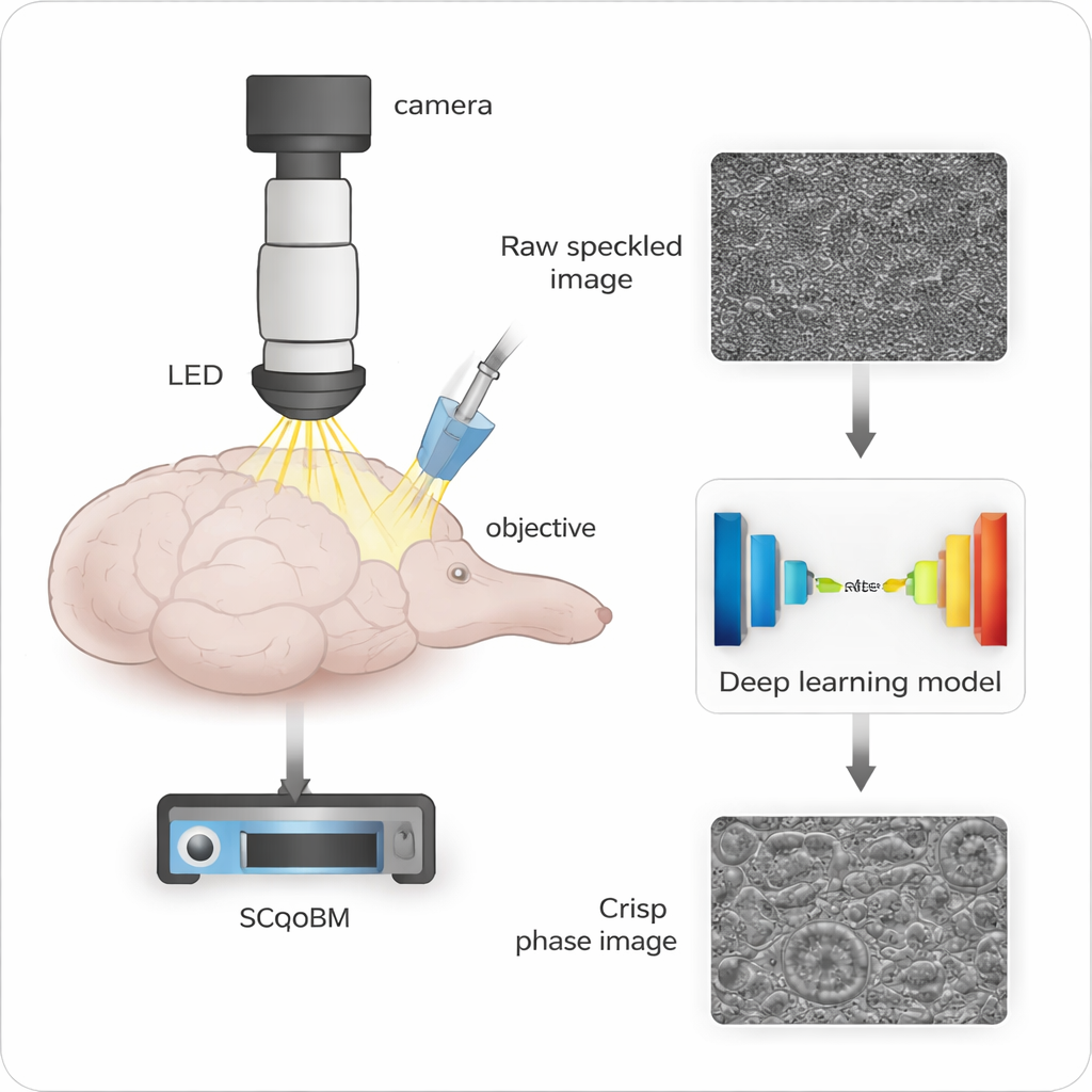
To remove this bottleneck, the authors created single-capture qOBM (SCqOBM). Instead of collecting four images from different directions, SCqOBM takes just one image with light shining in from a single oblique angle. A deep learning model—built on a U-Net, a popular image-processing neural network—then learns to convert this single raw picture into the same kind of detailed map that four images used to produce. The team trained and tested this network using thousands of examples where the “correct answer” was already known from standard four-capture qOBM, allowing the model to learn how subtle brightness patterns correspond to true tissue structure.
Proving It Works on Blood and Brain
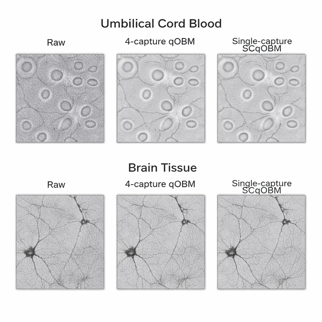
First, the researchers tested SCqOBM on umbilical cord blood stored in collection bags. Blood cells are relatively simple and symmetric, making them an ideal starting point. They showed that both single-capture and two-capture versions of the method reproduced the shapes and optical properties of red and white blood cells almost exactly, with only small numerical differences from the four-capture gold standard. In some cases, the single-capture method even produced cleaner images because it used a color of light less strongly absorbed by hemoglobin, which reduced noise in the measurements.
They then moved to a tougher challenge: thick rat brain tissue, including healthy cortex, tumors, and tumor margins. These samples have intricate and highly varied structures. Even here, the deep learning reconstructions closely matched traditional qOBM, capturing both coarse tumor regions and fine details in normal brain tissue. Remarkably, a model trained only on rat brain images also worked well on human brain tumor samples, suggesting that the approach generalizes across species and tissue types. Analysis in the frequency domain confirmed a subtle limitation: because SCqOBM only sees light from one angle, it cannot fully recover information along a narrow band of directions, but it does not “hallucinate” missing structures; it simply leaves that band slightly underrepresented.
Watching Blood Flow in Real Time
With its speed advantage, SCqOBM can capture rapid processes that would blur with multi-shot methods. The team used a high-speed camera to record mouse brain blood vessels at about 2,000 frames per second, then used the SCqOBM model to turn each frame into a quantitative map. By following how the refractive index pattern from flowing blood cells shifted over time, they measured flow speeds from about 1 millimeter per second in tiny vessels up to more than 60 millimeters per second in larger ones, matching expected blood flow profiles. They could even track slow, rolling white blood cells along vessel walls—events linked to immune responses and inflammation—as the animal’s condition changed.
Three-Dimensional Views of Human Skin
Finally, the authors showed that SCqOBM can capture volumetric images of living human skin on the arm, at near video rates. By rapidly stepping the focus up and down with a piezo stage, they collected stacks of single-capture images, converted each to phase using SCqOBM, and then refined the volume with a second deep learning algorithm. The resulting 3D views reveal distinct skin layers and tiny capillaries carrying individual red blood cells at depths of over 100 micrometers. Depending on how wide an area they image and how many depth slices they take, they can trade field of view for speed, reaching up to 10 volumes per second while maintaining cellular and subcellular detail.
What This Could Mean for Medicine
In simple terms, this work shows that a microscope can use a single flash of light and artificial intelligence to reconstruct rich, three-dimensional information from thick, living tissue, without dyes or physical contact. While there are still limits—for example, some directions of fine detail are harder to recover from just one lighting angle—the method delivers image quality close to slower, more complex systems, while reaching speeds comparable to the fastest light-sheet microscopes. Because the hardware is relatively straightforward—a brightfield microscope with a single LED—SCqOBM could eventually make advanced, label-free imaging more accessible in research labs and clinics, enabling non-invasive blood analysis, real-time monitoring of brain and skin, and other applications where speed and gentleness are critical.
Citation: Casteleiro Costa, P., Bharadwaj, S., Li, Z. et al. Single capture quantitative oblique back-illumination microscopy. npj Imaging 4, 13 (2026). https://doi.org/10.1038/s44303-026-00147-w
Keywords: label-free imaging, deep learning microscopy, quantitative phase imaging, blood flow measurement, in vivo skin and brain imaging