Clear Sky Science · en
pan-ASLM: Axially Swept Light Sheet Microscopy for Fast and High-Resolution Imaging of Expanded Samples
Seeing the Invisible Inside Cells
Modern biology is driven by a simple desire: to actually see what is going on inside cells and tissues, down to the tiniest structures that keep us alive. But as scientists push for ever finer detail over ever larger regions of organs and brains, traditional microscopes are hitting hard limits in speed and field of view. This paper introduces a new imaging system, called pan-ASLM, that lets researchers rapidly scan huge, physically enlarged biological samples while still resolving features tens of nanometers across—fine enough to distinguish details such as the internal folds of mitochondria or the tiny junctions between nerve cells.
Making Cells Bigger to See More
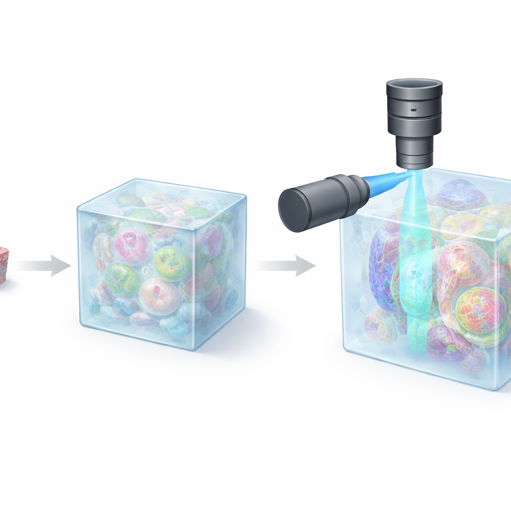
One of the most creative tricks in modern microscopy is to physically swell biological specimens. In “expansion microscopy,” cells or tissues are embedded in a special gel that soaks up water and expands uniformly, stretching all internal structures apart by a factor of roughly 4 to 20 in each dimension. The authors’ own variant, pan-ExM, can enlarge samples about 13–24 times while keeping most proteins in place and then labeling them with fluorescent dyes. Under a conventional light microscope, these swollen samples suddenly reveal details that previously required electron microscopes. But this success comes with a catch: after expansion, a once tiny region of tissue becomes enormous, turning routine three-dimensional imaging into a slow, data-heavy challenge.
Why Old Microscopes Fall Short
Standard confocal microscopes scan one point at a time and reject out-of-focus light through a pinhole, yielding crisp images but at the cost of speed and field of view. With expanded samples, signal levels are lower and more averaging is needed, so recording a single 3D stack over a modest area can take hours. Spinning-disk confocal systems parallelize the process and run faster, but they work best with high-magnification lenses that see only small regions and have short reach into the sample. Attempts to switch to wide-field objectives tend to sacrifice resolution, especially along the viewing axis, undermining the very gains expansion microscopy was meant to deliver.
A New Way of Lighting Up Tissues
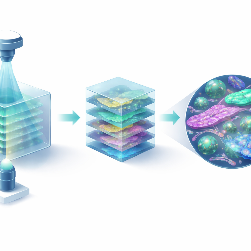
Light sheet fluorescence microscopy offers another path: it illuminates only a thin slice of the sample from the side, while a second lens collects the emitted light at right angles. This design naturally speeds up imaging and improves contrast, because most of the sample is kept dark. However, classical light sheets must balance how thin they are against how far they extend, forcing a tradeoff between sharpness and coverage. Axially swept light sheet microscopy (ASLM) tackles this by rapidly shifting a very thin sheet through the sample and matching that motion to the readout of a fast camera. In this work, the authors build pan-ASLM, an ASLM instrument tailored from the ground up for large, water-based expanded samples, using carefully chosen lenses, a calibrated high-speed voice coil to move the light sheet, and a wide, high-pixel-count camera.
Sharper, Faster Views of Cells and Organs
Put to the test, pan-ASLM delivers nearly equal clarity in all three dimensions, with effective resolutions of about 25–30 nanometers in expanded specimens. It images areas of 640 by 640 micrometers at up to 20 frames per second, achieving roughly 1200 times the imaging speed, seven times the field of view, and about twice the axial resolution of a typical confocal microscope used on similar samples. The team shows that this performance is not just a technical benchmark: in expanded human cells, they clearly resolve mitochondrial cristae, the layered components of nucleoli, and ring-like nuclear pores. In mouse kidney tissue, they capture fine brush borders and delicate foot processes in filtration units. In mouse brain cortex, they stitch together many tiles to reconstruct millimeter-scale volumes where individual synapses, the junctions between neurons, remain sharply defined regardless of their orientation.
Opening the Door to Big Biological Questions
By marrying sample expansion with a purpose-built light sheet microscope, pan-ASLM turns what used to be a painstaking, hours-long task into a practical, minutes-long measurement, without giving up nanoscale detail. This shift makes it realistic to map the architecture of organs, trace neural connections, or quantify the shapes and protein content of tiny structures across large tissue regions. As cameras, lasers, and dyes continue to improve, the authors foresee even larger, faster studies, paired with automated image analysis and machine learning. For non-specialists, the key message is that we are entering an era where scientists can routinely explore the inner landscape of cells and brains over vast areas with near-electron-microscope detail, using light-based tools that are finally fast and flexible enough to keep up.
Citation: Mekbib, H.T., Andersen, L.P., Zhang, S. et al. pan-ASLM: Axially Swept Light Sheet Microscopy for Fast and High-Resolution Imaging of Expanded Samples. npj Imaging 4, 16 (2026). https://doi.org/10.1038/s44303-026-00141-2
Keywords: expansion microscopy, light sheet imaging, super resolution, brain mapping, tissue ultrastructure