Clear Sky Science · en
Low molecular weight 4,4’-quinocyanines for in vivo NIR-II fluorescence imaging
Lighting Up Hidden Tumors
Surgeons increasingly rely on glowing dyes to spot cancers in real time, but today’s tools struggle to reveal tumors buried beneath the surface. This study introduces a new family of small, glowing molecules that shine in a deeper part of the infrared spectrum, allowing doctors to see farther into the body with sharper contrast and less background haze. If successfully translated to the clinic, these dyes could help surgeons remove more cancer while sparing healthy tissue.
Why Deeper Light Matters
Most clinical fluorescent dyes used today glow in the near‑infrared “NIR‑I” range, which already penetrates tissue better than visible light. Yet even these are limited by scattering and natural glow from our own tissues, making it difficult to clearly see structures more than a few millimeters deep. By shifting fluorescence into a longer‑wavelength region called “NIR‑II,” light scatters less and tissue gives off almost no background signal. The result is the potential for crisper images and the ability to look deeper into organs, blood vessels, and tumors during surgery.

Designing a New Family of Glow
The authors engineered a new class of organic dyes called 4,4'-quinocyanines (QuCy). Building on a well‑known surgical dye scaffold (cyanine dyes), they replaced one part of the molecule with a quinoline unit that extends the system of connected electrons. Computer calculations showed that this change narrows the energy gap between the molecule’s ground and excited states, which in turn pushes its color toward longer, NIR‑II wavelengths. Using a modular synthetic route, the team created both flexible and more rigid versions of QuCy dyes and fine‑tuned features like water-loving and fat-loving groups so the molecules could be formulated and linked to future targeting units such as peptides or antibodies.
Brighter, Smaller, and Built for the Body
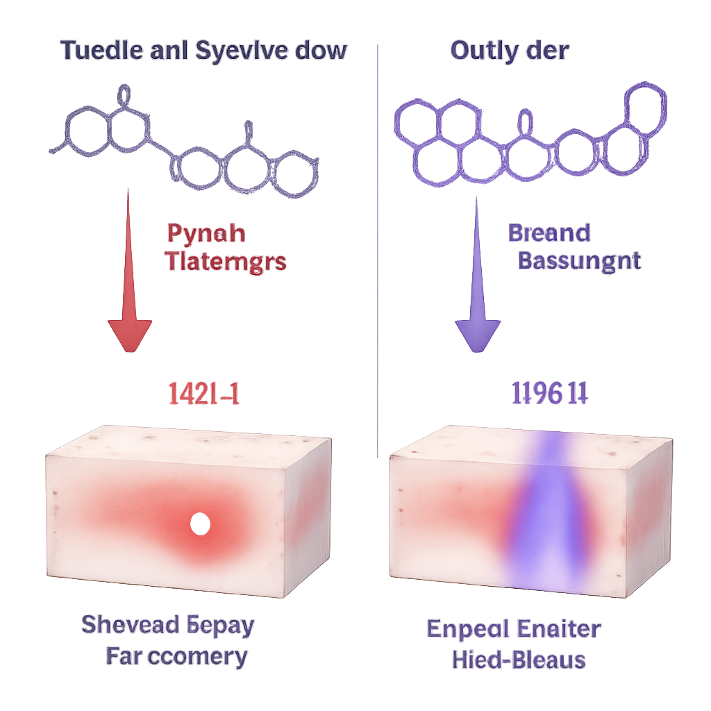
Laboratory measurements revealed that the new dyes absorb and emit light at much longer wavelengths than standard cyanines: absorption peaks near 940–970 nanometers and emission around 976–1004 nanometers, comfortably in the NIR‑II window. Importantly, these molecules are tiny—roughly half or less the size of many existing NIR‑II agents, which are often bulky polymers. Despite their compact size, several QuCy dyes were bright and stable under prolonged illumination, especially when packed into tiny fat‑like bubbles called liposomes. Experiments with tissue‑mimicking gels and slices of chicken breast showed that QuCy dyes maintained sharp, localized signals through up to 6 millimeters of tissue, whereas current NIR‑I dyes became fuzzy and lost most of their intensity beyond 2–3 millimeters.
From Cells to Living Mice
When tested on lung cancer cells in the lab, only some QuCy variants glowed strongly inside intact cells, revealing that both cell entry and the dye’s local environment matter for brightness. The cyclic QuCy dye named JAM317 stood out, giving robust intracellular fluorescence and remaining stable when packaged in liposomes. In live mice, JAM317 delivered high‑resolution images of the blood vessel network when illuminated in the NIR‑II range. Compared head‑to‑head with the commonly used surgical dye indocyanine green, JAM317 produced clearer vessel outlines and finer detail, particularly when detected at longer wavelengths. Tracking where the dye went over time showed rapid passage through the heart and lungs, followed by accumulation in the liver and eventual clearance through the intestines, consistent with strong binding to blood proteins and a primarily liver‑based route out of the body.
Toward Smarter Surgical Imaging
Overall, the study demonstrates that small, carefully engineered QuCy dyes can overcome key drawbacks of current fluorescent agents by offering deeper penetration, lower background, and high‑detail images, all in a compact, tunable package. For non‑specialists, the takeaway is that surgeons may soon have “night‑vision goggles” for cancer: injectable dyes that safely light up tumors and blood vessels deep inside the body, helping doctors see more, cut more precisely, and leave behind less disease.
Citation: Isuri, R.K., Hart, M.C., Adusei-Poku, S. et al. Low molecular weight 4,4’-quinocyanines for in vivo NIR-II fluorescence imaging. npj Imaging 4, 15 (2026). https://doi.org/10.1038/s44303-026-00140-3
Keywords: fluorescence-guided surgery, near-infrared imaging, NIR-II dyes, tumor visualization, vascular imaging