Clear Sky Science · en
A probabilistic deep learning approach for choroid plexus segmentation in autism spectrum disorder
Why this work matters for brain health and autism
The choroid plexus is a tiny structure deep inside the brain that helps produce and filter the fluid bathing our brain and spinal cord, and it also plays a key role in immune activity in the brain. Growing evidence suggests that in some people with autism spectrum disorder (ASD), this structure may look or behave differently, potentially reflecting changes in brain inflammation. To truly understand these links, scientists need to study thousands of brain scans—but doing so requires fast, reliable computer tools that can find and outline the choroid plexus automatically. This study introduces and tests such a tool, showing not just how well it works, but also how confident it is in its own answers.
A small but powerful brain gateway
The choroid plexus sits in the brain’s fluid-filled spaces and forms a barrier between blood and the clear liquid called cerebrospinal fluid. It helps control what enters and leaves the brain’s environment and is involved in immune signaling, including responses linked to inflammation. Earlier research has found that the choroid plexus can be enlarged or altered in several brain conditions, from multiple sclerosis to depression, and initial studies suggest differences may also be present in some autistic individuals. However, carefully tracing this structure by hand on MRI scans is slow, demanding, and somewhat subjective, which makes large-scale research nearly impossible without automation.
Teaching a computer to find the choroid plexus
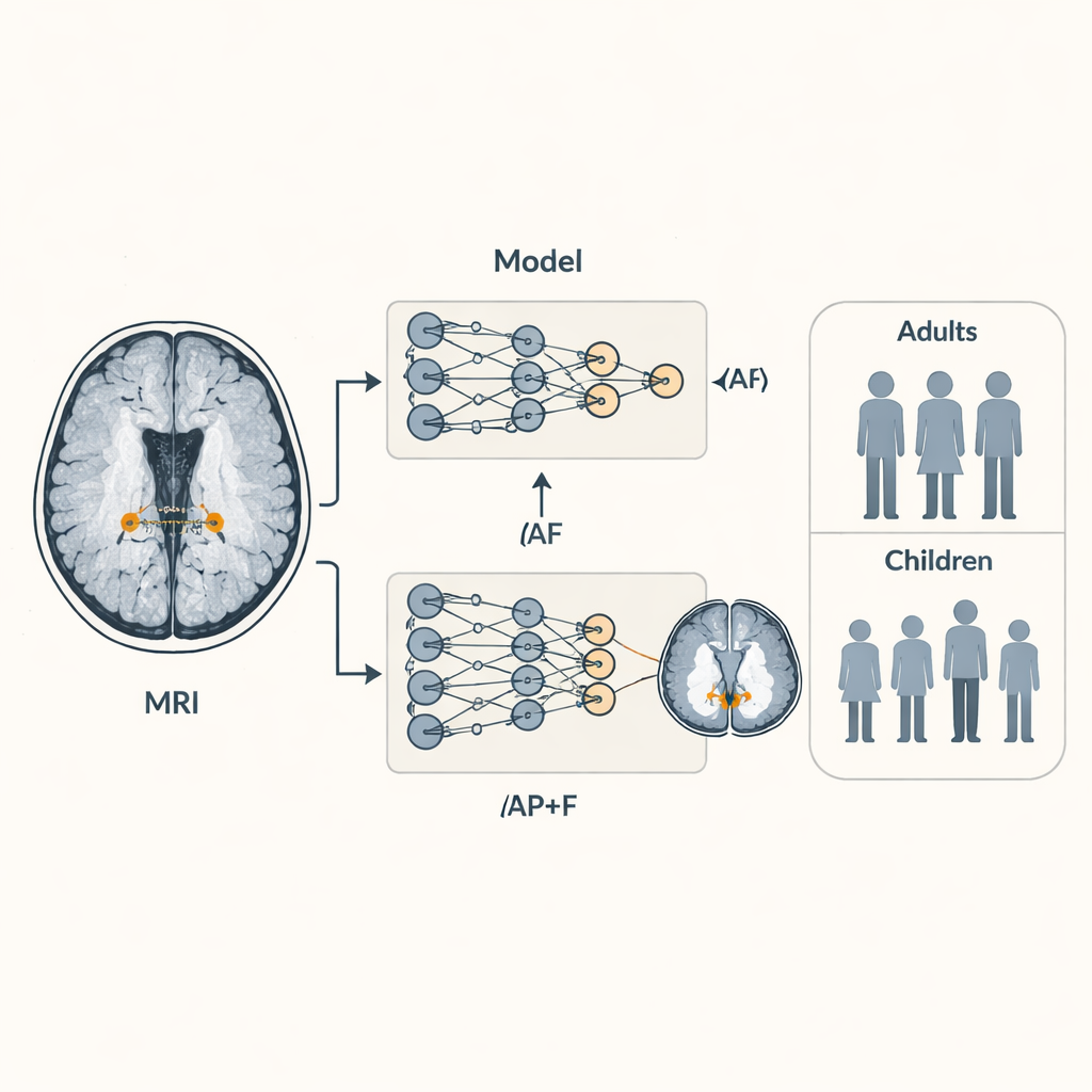
The authors focused on ASCHOPLEX, a recently developed deep learning system that automatically segments, or outlines, the choroid plexus on MRI scans. Originally trained on adults with and without multiple sclerosis, ASCHOPLEX had already shown near-human accuracy in other groups. In this study, the team adapted the tool for ASD by “finetuning” it using a small but carefully labeled set of 12 adults (with and without autism) from a local research project. They then tested how well it worked on an additional 53 adults whose choroid plexus had been traced manually by experts, allowing a direct comparison between human and machine. They also compared ASCHOPLEX to a widely used brain MRI tool called FreeSurfer, which was not specifically designed for this structure.
Adding a sense of confidence to the predictions
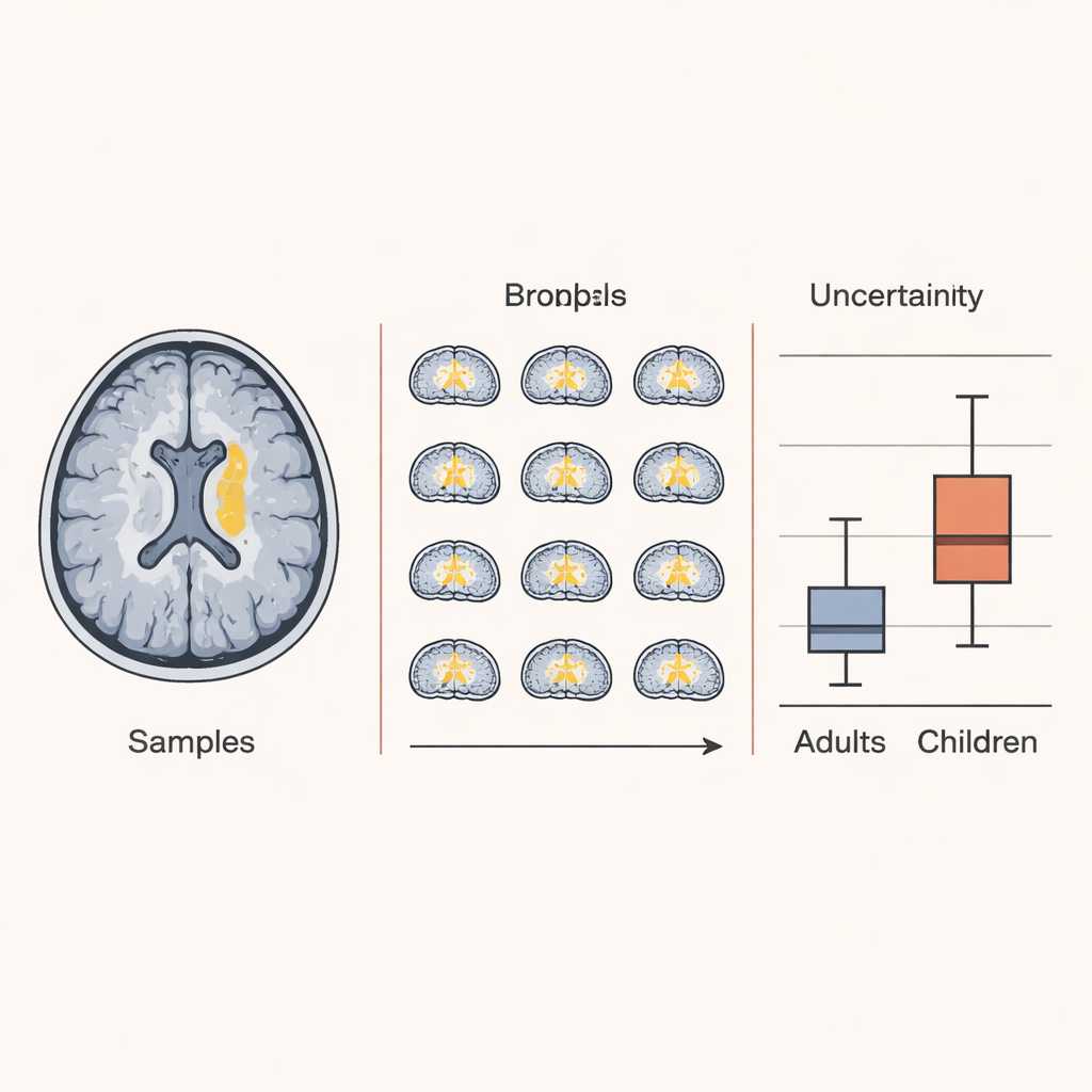
Beyond simply asking whether the tool was right or wrong, the researchers wanted to know how sure it was about each decision. To do this, they turned ASCHOPLEX into a “probabilistic” model by switching on a technique called dropout during both training and testing. In practical terms, this means the model is run many times on the same scan, each time with slightly different internal settings, producing a collection of slightly different predictions. By looking at how much those predictions agree or disagree at each point in the brain, the team could estimate uncertainty—places where the model is confident and places where it is not. They applied this approach not only to their local adult dataset, but also to more than 1,800 participants, children and adults, from the large Autism Brain Imaging Data Exchange (ABIDE) project.
How well the tool worked across people and ages
After finetuning, ASCHOPLEX closely matched human-drawn outlines of the choroid plexus in adults with and without autism, achieving accuracy levels similar to or better than agreement between human experts. It clearly outperformed FreeSurfer, which was never optimized for this structure. Importantly, once finetuned, ASCHOPLEX no longer showed performance differences between autistic and non-autistic adults or between men and women, reducing concerns about systematic bias. When the probabilistic version was used on the large ABIDE dataset, the model remained most confident for adults, especially those resembling the training group, but its uncertainty rose for both adults and children from outside sites—and was highest in children. Detailed analysis showed that this extra uncertainty mainly reflected the model’s lack of familiarity with children’s brain scans, rather than poor scan quality.
What this means for future autism research
For non-specialists, the key message is that researchers now have a practical AI-based tool that can accurately find a very small, important brain structure in people with and without autism, and can say how sure it is about each result. ASCHOPLEX, especially in its probabilistic form, can be applied to large imaging collections to screen for choroid plexus changes that might signal altered immune activity in the brain. At the same time, its rising uncertainty in children highlights that such tools still need additional training on younger populations before they can be fully trusted in all age groups. Overall, the study shows how combining deep learning with explicit confidence measures can make brain imaging analyses both more powerful and more transparent, paving the way for better understanding of neuroimmune changes in autism.
Citation: Bargagna, F., Morin, T.M., Chen, YC. et al. A probabilistic deep learning approach for choroid plexus segmentation in autism spectrum disorder. NPP—Digit Psychiatry Neurosci 4, 2 (2026). https://doi.org/10.1038/s44277-026-00056-1
Keywords: autism spectrum disorder, choroid plexus, brain MRI, deep learning, neuroinflammation