Clear Sky Science · en
In vivo acoustoelectric neural recording in mice enabled by ultrasound-induced frequency mixing
Listening to the Brain Without Opening the Skull
Diagnosing and treating brain disorders often requires eavesdropping on the brain’s tiny electrical whispers. Today, doctors must choose between non-invasive methods that blur together large regions of the brain, or invasive implants that require surgery. This study introduces a new approach in mice that borrows tricks from radio engineering and medical ultrasound, hinting at future scanners that could “tune in” to deep brain activity without opening the skull.
Why Current Brain Scans Are Not Enough
Common tools to measure brain activity each come with trade-offs. Electroencephalography (EEG) listens to the brain’s electrical activity through sensors on the scalp, but the skull smears and weakens the signals, so only large, surface-wide events can be seen clearly. Magnetoencephalography (MEG) can pinpoint activity more precisely but mainly near the outer layers of the brain. Functional MRI offers three-dimensional images but does not measure electrical activity directly; instead, it tracks slow changes in blood flow. None of these methods can non-invasively pick out fast, tiny electrical changes from a small, deep patch of brain tissue with high precision.
Using Sound Waves to Focus on Tiny Brain Regions
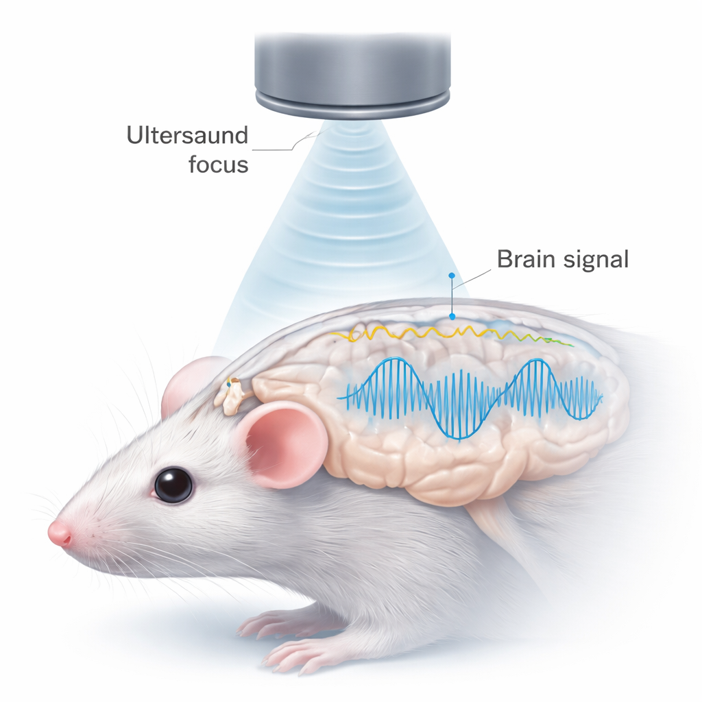
Ultrasound—the same kind of sound used in prenatal scans—can be focused like a spotlight into the body, including at depths inside the skull when distortions are corrected. The authors take advantage of a physical effect called the “acoustoelectric” interaction: when sound waves pass through salty tissue carrying an electrical signal, the two can mix. In essence, the local brain signal at the ultrasound focus rides on top of a high-frequency sound “carrier,” much like a radio station rides on a radio wave. This mixing shifts the brain’s low-frequency electrical activity up to much higher frequencies, where it can be separated from background noise and other brain signals using standard demodulation techniques from radio engineering.
Testing the Idea in Salty Water and Mouse Brains
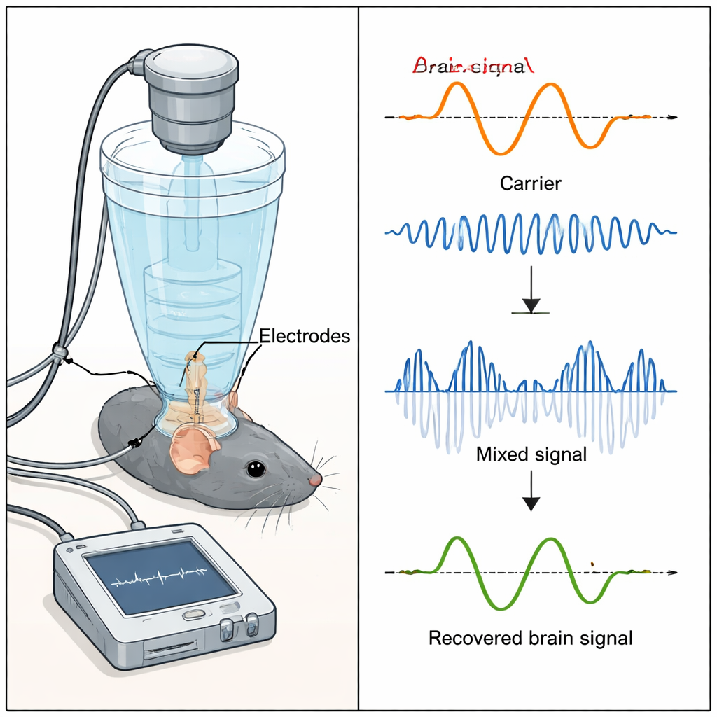
To check that this mixing really happens and is not just a recording glitch, the team first used a dish of salty water with tiny electrodes and a focused ultrasound beam. They showed that only where the ultrasound was focused did the expected “sum and difference” frequencies appear around the carrier, confirming true local mixing rather than simple electrical interference. They then refined their signal processing, using special spectral windows and narrow frequency bands, to tease out extremely small mixed signals—similar in size to real brain signals—from beneath large artefacts caused by the ultrasound hardware itself.
Reading Vision Signals and Spontaneous Activity
Next, the researchers implanted fine electrodes in the visual cortex and motor cortex of mice. While lightly anesthetized, the mice viewed a green light flashing at 8–10 times per second, which evokes a well-known rhythmic brain response in visual areas. At the same time, the team continuously applied focused ultrasound at 500 kHz. They showed that the usual visual brain signal could still be measured in the normal, low-frequency range, even during ultrasound, meaning the method did not drown out ordinary recordings. Crucially, by filtering the data only around the ultrasound frequency and then demodulating it, they were able to reconstruct a version of the original visual response from the mixed, high-frequency signal alone. They further demonstrated that this reconstruction depended on the presence of the acoustic field and on tuning to the correct carrier frequency, ruling out simple electrical crosstalk.
Toward Real-Time, Non-Invasive Brain Listening
Finally, the authors showed that they could recover spontaneous, non-repeated brain activity from single trials—not just averaged responses to repeated flashes. This suggests that, in principle, acoustoelectric neural recording could one day provide real-time monitoring of ongoing brain activity with a spatial precision set by the ultrasound focus rather than by electrode placement. Important challenges remain, especially safely delivering and detecting such small mixed signals through the thicker human skull and managing heating from continuous ultrasound. Still, this proof-of-concept in mice outlines a path toward portable, non-invasive devices that could listen to local brain circuits using focused sound, offering new ways to study and perhaps diagnose conditions like epilepsy, depression, and other brain disorders.
Citation: Rintoul, J.L., Howard, J., Dzialecka, P. et al. In vivo acoustoelectric neural recording in mice enabled by ultrasound-induced frequency mixing. Commun Eng 5, 37 (2026). https://doi.org/10.1038/s44172-026-00598-4
Keywords: ultrasound brain imaging, noninvasive neural recording, acoustoelectric effect, visual evoked potentials, brain signal decoding