Clear Sky Science · en
Evaluating flow modulating treatment response in intracranial aneurysms using black-blood MRI in vitro
Seeing Dangerous Brain Bulges in a New Light
Brain aneurysms—balloon-like bulges in blood vessels—can rupture without warning and cause life-threatening bleeding. Doctors increasingly treat them from the inside using tiny mesh implants that calm the blood flow and help the bulge seal off. But right after such a device is placed, it is surprisingly hard to measure whether blood flow has actually slowed enough for the treatment to succeed. This study explores whether a widely available MRI technique, called black-blood MRI, can act as a simple visual gauge of how well these devices are working, potentially reducing the need for more invasive imaging.
Why Blood Flow Matters for Healing
Aneurysm implants such as flow-diverting stents and intrasaccular devices are designed to reroute blood so that less of it rushes into the weakened bulge. When flow inside the aneurysm drops, a stable clot can form and the vessel lining can slowly grow over the opening, eventually closing off the aneurysm. Previous work has shown that if the blood slows only a little, the aneurysm may continue to fill, grow, or even rupture despite treatment. Today, doctors mostly rely on X-ray angiography, which requires injecting contrast dye and exposes patients to radiation, or on a specialized MRI method called 4D flow MRI, which directly measures blood velocity but is slow and easily disturbed by metal from the implants. The authors asked whether black-blood MRI—usually used to make vessel walls stand out—could indirectly reveal where blood has slowed after treatment.
Building Brain Vessel Models in the Lab
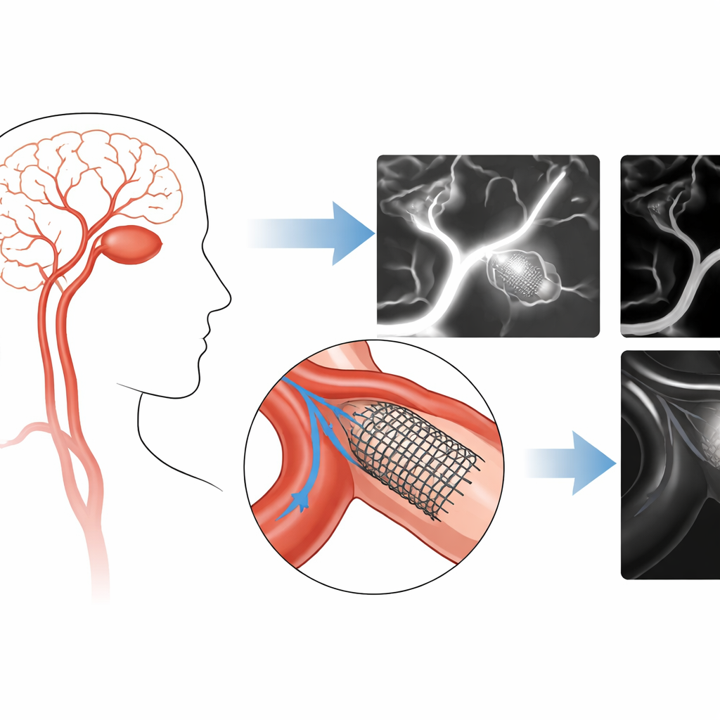
To test this idea under controlled conditions, the researchers created highly detailed 3D-printed models of brain arteries from real patient scans: one with an aneurysm on the internal carotid artery and several basilar artery models with different aneurysm sizes. These plastic replicas were connected to pumps that drove blood-like fluids through them in a closed loop, mimicking realistic heartbeats. The team placed commercial and prototype implants into the models—tube-shaped flow-diverter stents spanning the aneurysm neck and basket-like intrasaccular devices filling the bulge. They then scanned all models with both 4D flow MRI, which provided direct blood-velocity measurements, and spin-echo “black-blood” MRI, in which fast-flowing blood appears dark and slower blood appears brighter.
Watching Flow Slow Down After Treatment
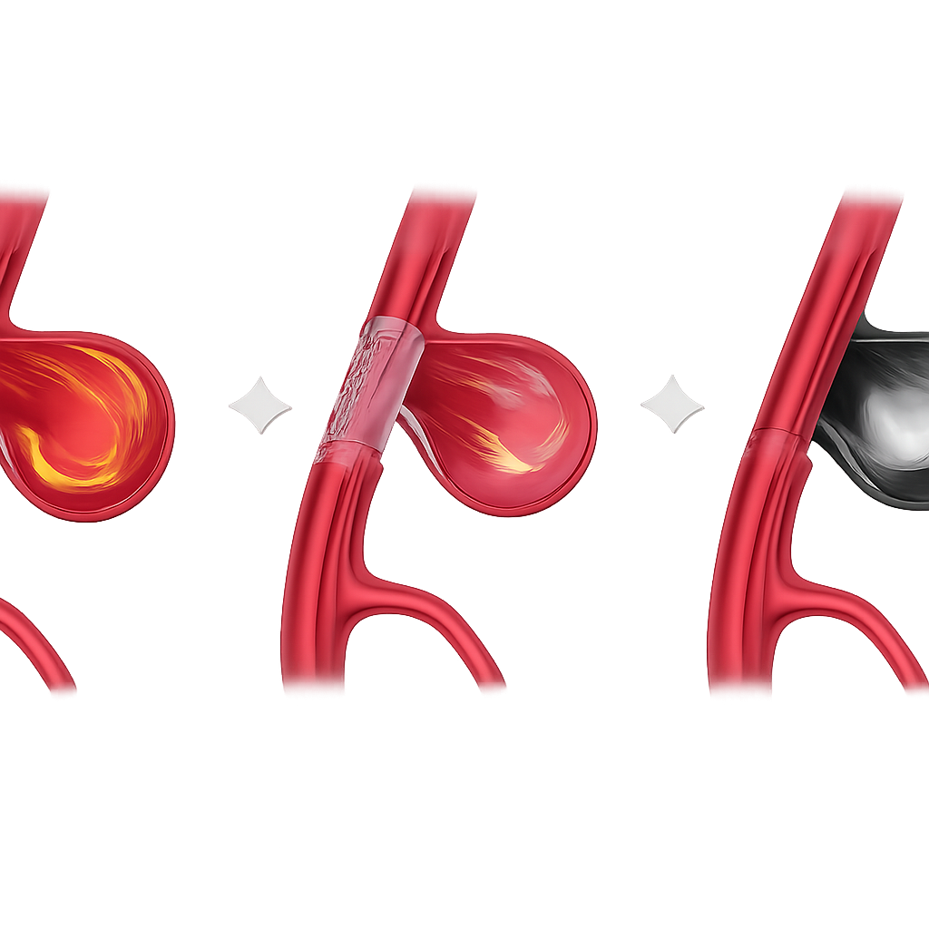
In straight tube models, the team first confirmed a basic relationship: as flow speed increased, the black-blood MRI signal decreased, and vice versa. This established that the technique is sensitive to flow speed. In the aneurysm models, the implanted devices behaved as intended: 4D flow MRI and computer simulations showed that blood speed inside the bulge dropped sharply after treatment, while the flow in the main parent vessel changed little. At the same time, black-blood MRI showed the opposite pattern in brightness: the aneurysm sac became markedly brighter after device placement, but the parent artery’s appearance stayed nearly the same. Across many devices and geometries, higher black-blood signal in the aneurysm consistently lined up with lower measured flow, revealing a strong inverse relationship between the two.
Working Around Metal and Other Real-World Challenges
A major practical advantage of the black-blood method emerged when strong metal artifacts appeared on 4D flow MRI, especially around the dense intrasaccular devices. In some experiments, the metal erased the MRI signal within the aneurysm region so severely that direct flow measurements were impossible. Black-blood MRI, however, was much less affected, still revealing most of the aneurysm and clearly showing a bright region where flow had slowed. Computer simulations confirmed that these bright areas matched zones of reduced velocity. The pattern held across different device designs, aneurysm shapes and sizes, and even different fluid viscosities, suggesting the approach is robust as long as scan settings are kept consistent.
What This Could Mean for Patients
To a non-specialist, the key takeaway is that a brighter spot in a treated aneurysm on black-blood MRI likely signals slow, stagnant blood—exactly what doctors hope to achieve after placing a flow-modulating implant. Because this scan is already used in many hospitals to look at vessel walls, it could double as a quick check of whether treatment has effectively tamed the blood flow, especially when metal devices make other MRI methods unreliable. The study was done in lab-grown models, not patients, so more clinical work is needed to translate brightness levels into firm rules for success or failure. Still, the findings suggest that a familiar imaging tool could be repurposed into a non-invasive, three-dimensional indicator of treatment response, helping doctors track which aneurysms are on a safe path to healing.
Citation: Pravdivtseva, M.S., Toraman, H., Korte, J. et al. Evaluating flow modulating treatment response in intracranial aneurysms using black-blood MRI in vitro. Commun Med 6, 170 (2026). https://doi.org/10.1038/s43856-026-01413-z
Keywords: brain aneurysm, MRI, blood flow, endovascular stent, medical imaging