Clear Sky Science · en
Motor learning induces myelin-related white matter changes revealed by MRI-based in vivo histology
How Practice Can Reshape the Wiring of the Brain
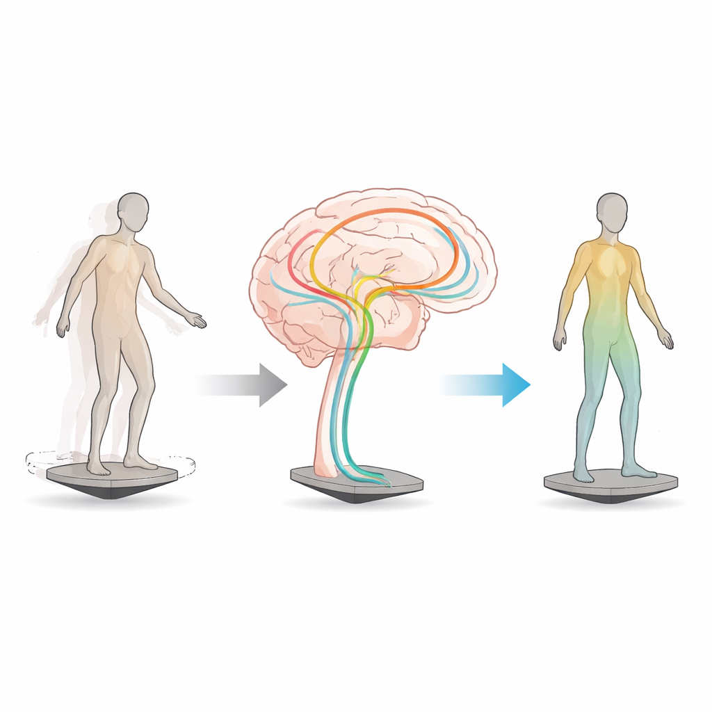
Learning to stay steady on a wobbling board may not sound like brain science, but it turns out that simple balance training can subtly reshape the brain’s internal wiring. This study asked a basic yet far-reaching question: when adults learn a new motor skill, such as keeping their balance on an unstable platform, how does the "white matter"—the long nerve cables that connect distant brain areas—actually change? Using advanced MRI scans, the researchers tracked these changes over weeks, revealing how practice can tune the brain’s communication highways in ways that may matter for learning, healthy aging, and rehabilitation.
Peeking Inside the Brain’s Wiring
Most people have heard that learning changes "gray matter," the regions packed with nerve cell bodies. But gray matter is only half the story. White matter, made of fiber bundles coated in fatty insulation, helps coordinate signals across the brain with split-second timing. Until recently, scientists could gauge white matter health only in broad strokes, without knowing which microscopic features were changing. In this study, 24 young adults first went through a four-week period with no training, then practiced a demanding whole-body balance task for another four weeks. At three points—before, during, and after this training—the researchers collected a battery of quantitative MRI scans designed to tease apart different features of brain tissue, such as fiber density, surrounding water, and properties linked to myelin, the insulating sheath around nerve fibers.
Following the Brain’s Motor Highways
Rather than inspecting the brain voxel by voxel, the team focused on specific white matter pathways that form the core of the motor network. Using diffusion-based tractography, they digitally "dissected" fiber bundles such as the corticospinal tracts that run from the motor cortex to the spinal cord, the fronto-pontine fibers that bridge the cortex and cerebellum, and thalamic pathways that relay signals between deep brain hubs and the frontal lobes. They then projected multiple MRI-derived measures onto many small segments along each bundle. To make sense of this rich, multicolored dataset, the researchers applied a multivariate method that looks for latent patterns of change over time—combinations of measures that tend to rise or fall together—rather than examining each MRI metric in isolation.
Practice-Linked Changes, Not Just Random Fluctuations
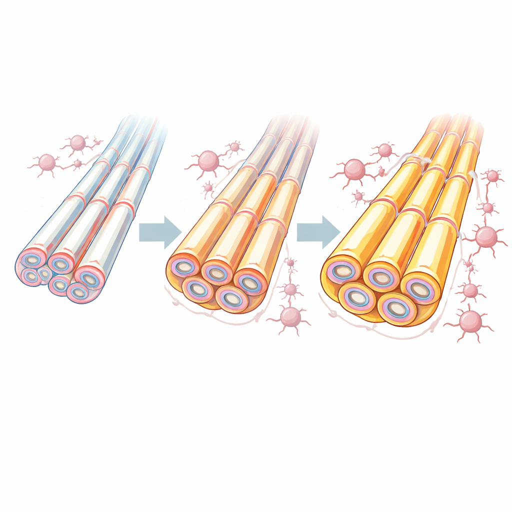
Across thousands of tract segments, only a small, coherent set showed changes that passed several strict tests. In five key locations—within the anterior thalamic radiation, thalamo–premotor pathway, fronto-pontine fibers, and both left and right corticospinal tracts—MRI patterns shifted during the training phase, but remained stable during the no-training period. The extent of these changes tracked how quickly individuals improved on the balance task, tying the brain alterations directly to learning rather than to the mere passage of time. In some regions, the dominant signal suggested reduced free water and increased tissue density, consistent with tighter packing or growth of support cells. In others, a composite measure called the aggregate g-ratio, thought to reflect the balance between fiber core and insulating sheath, shifted in a direction compatible with enhanced myelination around axons.
A Coordinated Brain-Wide Response
Intriguingly, these learning-related modifications did not behave like independent, isolated tweaks. When the researchers summarized the main pattern of change in each of the five segments and looked at how these summaries related to one another, they found that a single underlying dimension explained most of the variation. In other words, when one part of the motor network’s wiring changed, other parts tended to change in concert, hinting at a network-wide adjustment rather than scattered, unrelated updates. This shared white matter plasticity also related to previously measured shifts in the fine structure of the overlying cortex in the same participants, supporting the idea that gray and white matter remodel together as new skills are acquired.
Why This Matters for Health and Rehabilitation
For non-specialists, the key message is that practicing a physical skill does more than strengthen muscles or refine reflexes—it also fine-tunes the hidden cables that link brain regions, possibly by adjusting their insulation and the surrounding support tissue. The study showcases a powerful way to combine several advanced MRI techniques to obtain a more biologically grounded picture of how white matter changes in living humans. Although the sample was modest and the exact cellular mechanisms remain partly inferred, the approach offers a roadmap for studying how training, aging, disease, or therapy reshape the brain’s wiring. In the future, such methods could help design and monitor interventions that harness white matter plasticity to improve movement, recovery after injury, or even everyday learning.
Citation: Aye, N., Kaufmann, J., Heinze, HJ. et al. Motor learning induces myelin-related white matter changes revealed by MRI-based in vivo histology. Commun Biol 9, 380 (2026). https://doi.org/10.1038/s42003-026-09712-w
Keywords: motor learning, white matter, myelin, brain plasticity, quantitative MRI