Clear Sky Science · en
ZIKV envelope protein is a strong blocker of early directional differentiation in the neural lineage
Why this matters for growing brains
Zika virus first hit headlines for causing babies to be born with unusually small heads and serious brain damage. But how does an infection in the mother’s body so powerfully derail the earliest steps of brain building in the embryo? This study zooms in on a single viral component – the envelope protein that coats Zika virus particles – and asks whether that protein alone can misguide the formation of nerve cells. By recreating early brain development in the lab with mouse stem cells, the researchers uncover how this viral protein quietly but strongly blocks the normal construction of neural circuits.
From flexible starter cells to future nerve cells
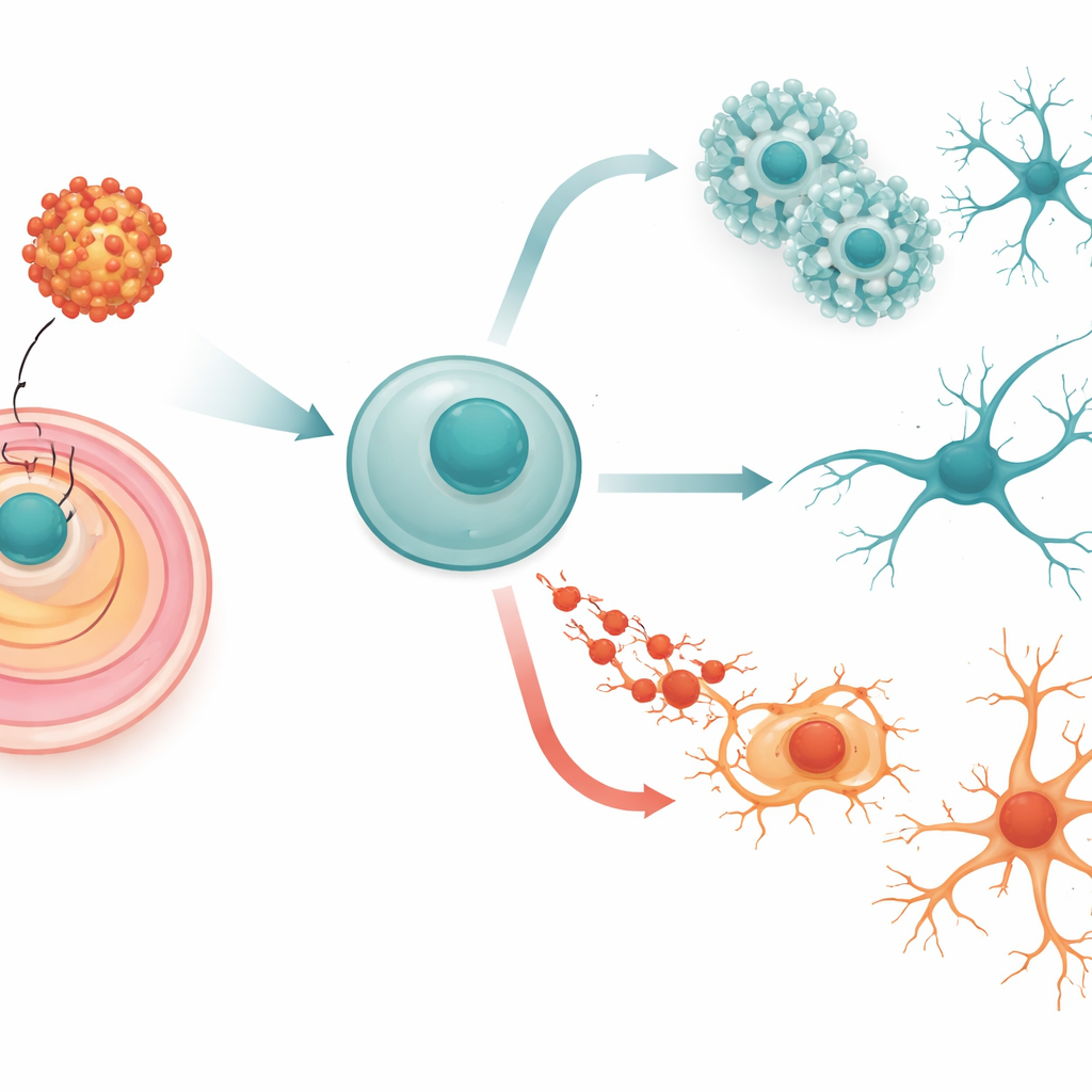
Our brains begin as simple, highly flexible cells called embryonic stem cells. These cells can turn into any tissue in the body, but under the right conditions they follow a carefully choreographed path toward becoming nerve cells. First they commit to the “neural” route, then form rosette-like structures that resemble the early nervous system, and finally mature into neurons that connect via long, branching processes. The team used mouse embryonic stem cells as a stand-in for these early stages and engineered them to produce the Zika envelope protein, with or without a small change at a key sugar attachment site known to affect the virus’s strength.
Viral coat protein freezes the first steps of brain building
When the stem cells made the Zika envelope protein, they still looked healthy and kept their broad potential to become many tissues. However, when allowed to differentiate freely, their ability to form complex 3D clusters representing the three basic tissue layers of the embryo was sharply reduced, and markers of all three layers dropped. This suggested that the viral protein does not kill the stem cells outright, but subtly interferes with their capacity to embark on normal developmental paths. A mutant version of the protein, lacking a specific sugar attachment, changed this pattern in a more uneven way, hinting that the fine chemical decoration of the protein tunes how it harms development.
Blocking the road from stem cell to neuron
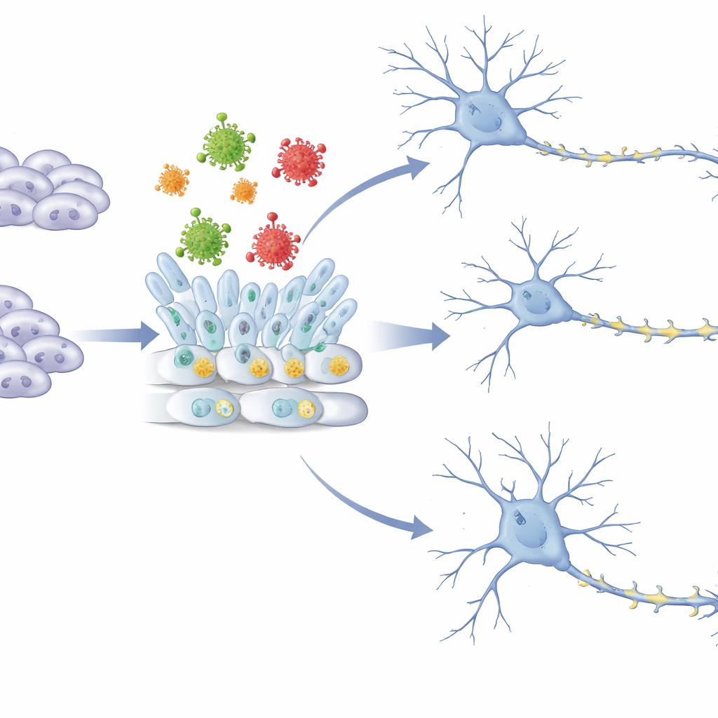
The researchers then focused specifically on the journey from stem cell to early neuron using two established lab models: a flat “monolayer” culture and a 3D “neurosphere” culture that mimics early brain tissue. In both systems, control cells smoothly increased their expression of neural stem and neuron markers over days, forming organized rosettes and abundant young neurons. Cells making the envelope protein, in contrast, produced far fewer neural stem cells, fewer rosettes, and many fewer early neurons, as seen by reduced levels of key genes and proteins linked to nerve identity. The sugar-free mutant form generally caused an even stronger block at the gene level and triggered additional inflammatory cell-death pathways, suggesting a route to more severe damage.
Silencing communication in young nerve networks
To understand what was going wrong inside the cells, the team compared global gene activity in normal and envelope-producing cells at critical differentiation stages. They found that many genes tied to nerve growth, synapse formation, and the tiny spines on dendrites that store memories were turned down. Pathways involved in loading and releasing neurotransmitters, wiring axons to their targets, and assembling synapses were all suppressed. At the same time, signaling routes linked to calcium and certain cell-surface receptors were switched on, potentially making cells overly excitable or mis-signaled. These sweeping changes appeared in both flat and 3D cultures, showing that the envelope protein repeatedly steers developing neural cells away from building robust, well-connected networks.
What this means for Zika-related birth defects
For non-specialists, the key message is that the Zika virus does not need to be actively multiplying and killing cells to harm the developing brain. This work shows that its outer coat protein alone can push early stem cells off the normal pathway toward becoming neurons, and can weaken the genetic programs needed to form healthy synapses and dendritic spines. Such early, quiet disruptions help explain how exposure in the womb can lead to conditions like microcephaly and long-term cognitive problems. The findings also warn that vaccines or therapies involving viral envelope proteins must be carefully assessed for potential effects on brain development, even when no live virus is present.
Citation: Ma, ZH., Wang, Y., Hassaan, N.A. et al. ZIKV envelope protein is a strong blocker of early directional differentiation in the neural lineage. Commun Biol 9, 395 (2026). https://doi.org/10.1038/s42003-026-09672-1
Keywords: Zika virus, brain development, neural stem cells, viral envelope protein, microcephaly