Clear Sky Science · en
enGLOW 3D microscopy of the enteric nervous system in cleared human and mouse gut
Seeing the Hidden Nerves of the Gut
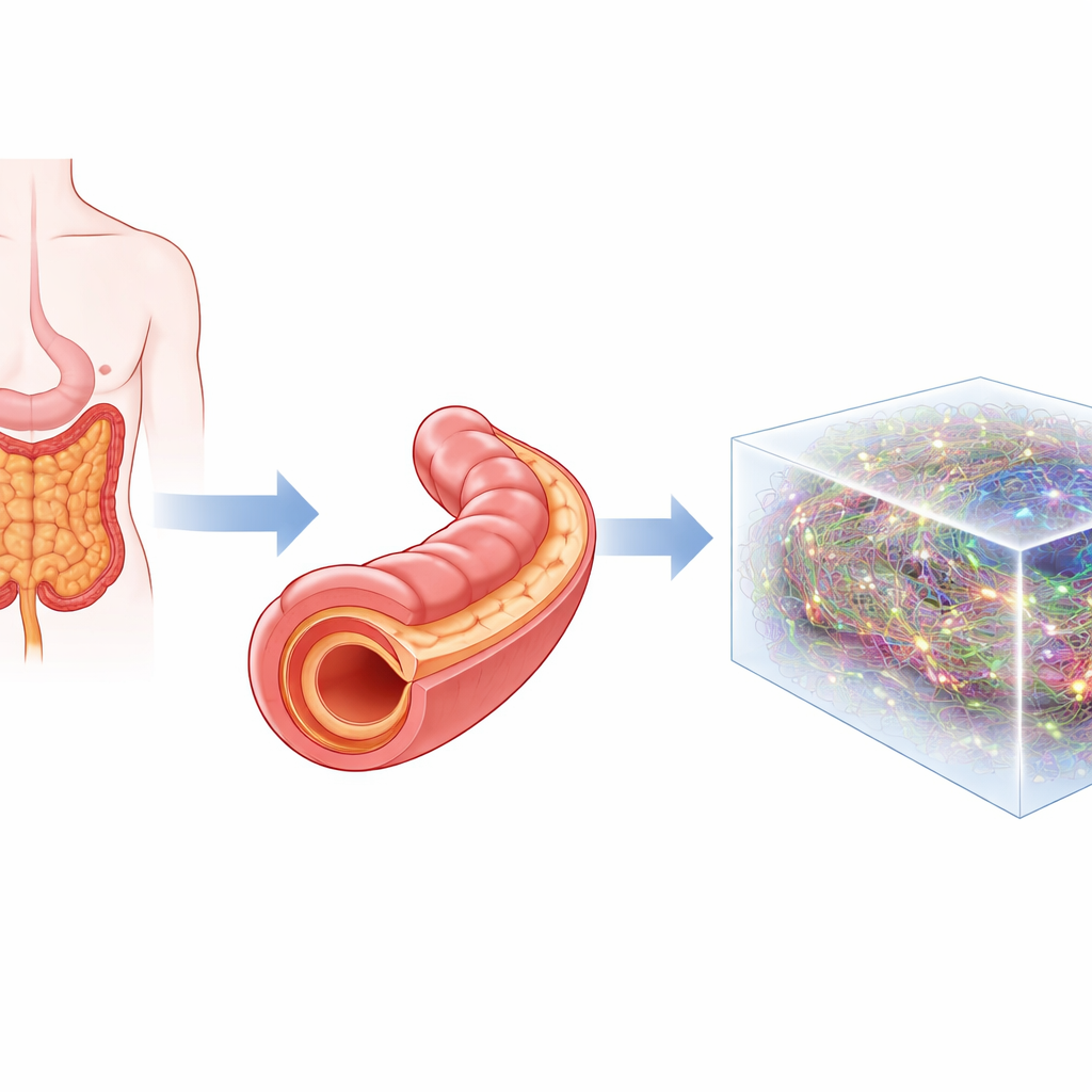
The gut is sometimes called our “second brain” because it holds its own vast nerve network that helps control digestion, immunity, and even links to the brain. Yet most of this wiring is buried deep in the wall of the intestine and has been notoriously hard to see as a whole. This paper introduces a new way to turn pieces of human and mouse gut into clear, three-dimensional samples so that researchers can map this hidden nerve network across large stretches of tissue, rather than in tiny slices.
A New Window into the Gut’s Nerve Network
The authors present enGLOW, a step-by-step laboratory workflow designed specifically for the gut. It combines chemical “clearing,” which makes intact tissue transparent, with light-sheet microscopes that scan large volumes in 3D. At the same time, the tissue’s own faint glow and added fluorescent tags are captured, revealing both the overall anatomy of the gut wall and the exact positions of different cell types. Unlike traditional methods that cut tissue into thin slices or peel off single layers, enGLOW keeps centimeter-scale pieces intact, allowing the entire local nerve network to be seen at once.
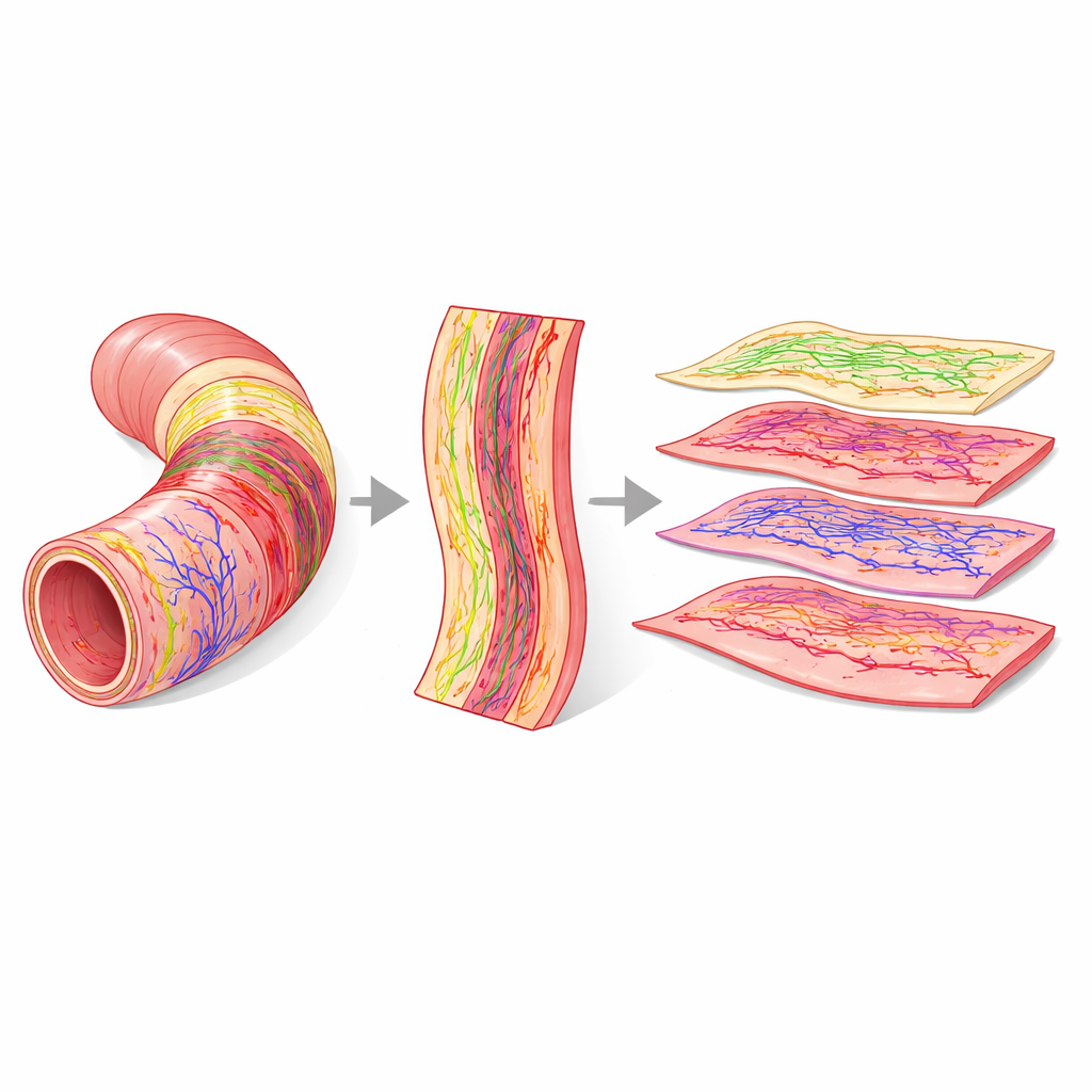
Turning Tubes into Flat Maps
One of enGLOW’s key innovations is a form of digital “virtual dissection.” The gut wall is made of several layers, including two nerve-rich sheets known as the myenteric and submucosal plexuses. In curved, tube-shaped tissue, these layers are hard to examine in full. The researchers use the gut’s outer surface as a reference and apply a computer algorithm that mathematically flattens the 3D images. This produces flat, layer-by-layer views of the same piece of tissue, separating nerve plexuses and muscle layers without physically cutting them. With this approach, they can compare how nerve cell clusters and fibers are arranged along different regions of the mouse digestive tract and measure how deeply each plexus lies beneath the surface.
Mapping Supporting Cells and Rhythm-Makers
Beyond nerve cells themselves, gut function depends on several partner cell types. Using enGLOW, the team labeled and imaged four major players in the mouse colon: nerve cell bodies, the long fibers that connect them, glial cells that support and regulate nerve activity, and interstitial cells of Cajal, which act as built-in pacemakers for gut movement. The 3D data, combined with virtual flattening, show how these cell networks weave through different layers of the gut wall, how densely they fill each region, and where they overlap or remain distinct. For example, the pacemaker cells form grid-like patterns aligned with muscle layers, while glia and nerve fibers spread broadly across several layers. This level of detail lets researchers quantify how much of a given layer is occupied by each network, not just whether the cells are present.
From Healthy Tissue to Disease Models
The workflow was also adapted for thick samples from human colon surgery. After clearing and embedding to preserve delicate layers, light-sheet imaging captured large blocks of human gut with enough resolution to see individual nerve clusters and the branching blood vessels they wrap around. In a mouse model of Parkinson’s disease, enGLOW revealed changes in the architecture of the gut lining and unusual patterns of nerve labeling in the mucosa, hinting at barrier disruption. Although the small number of animals prevents firm conclusions, these examples show how the method can uncover subtle structural changes that may accompany neurological disorders and other diseases tied to the gut.
Why This Matters for Health and Disease
For a layperson, the key message is that we now have a way to see the gut’s nerve “wiring diagram” across large, intact pieces of tissue, in both animals and humans. enGLOW turns what used to be fragmented snapshots into full 3D maps, and then digitally peels apart the gut wall to inspect each layer in turn. This makes it possible to measure how nerve networks, support cells, and pacemaker cells are organized, and how they remodel in conditions such as inflammatory bowel disease, diabetes, Hirschsprung’s disease, or Parkinson’s disease. Over time, such detailed maps of gut structure may help connect symptoms to specific changes in tissue architecture and guide new therapies that target the gut’s own nervous system.
Citation: Planchette, A., Gantar, I., Scholler, J. et al. enGLOW 3D microscopy of the enteric nervous system in cleared human and mouse gut. Commun Biol 9, 357 (2026). https://doi.org/10.1038/s42003-026-09643-6
Keywords: enteric nervous system, 3D gut imaging, tissue clearing, light-sheet microscopy, gut–brain axis