Clear Sky Science · en
Microstructural variation of hippocampal substructures across childhood and adolescence quantified with high-gradient diffusion MRI
Why this brain area matters as kids grow
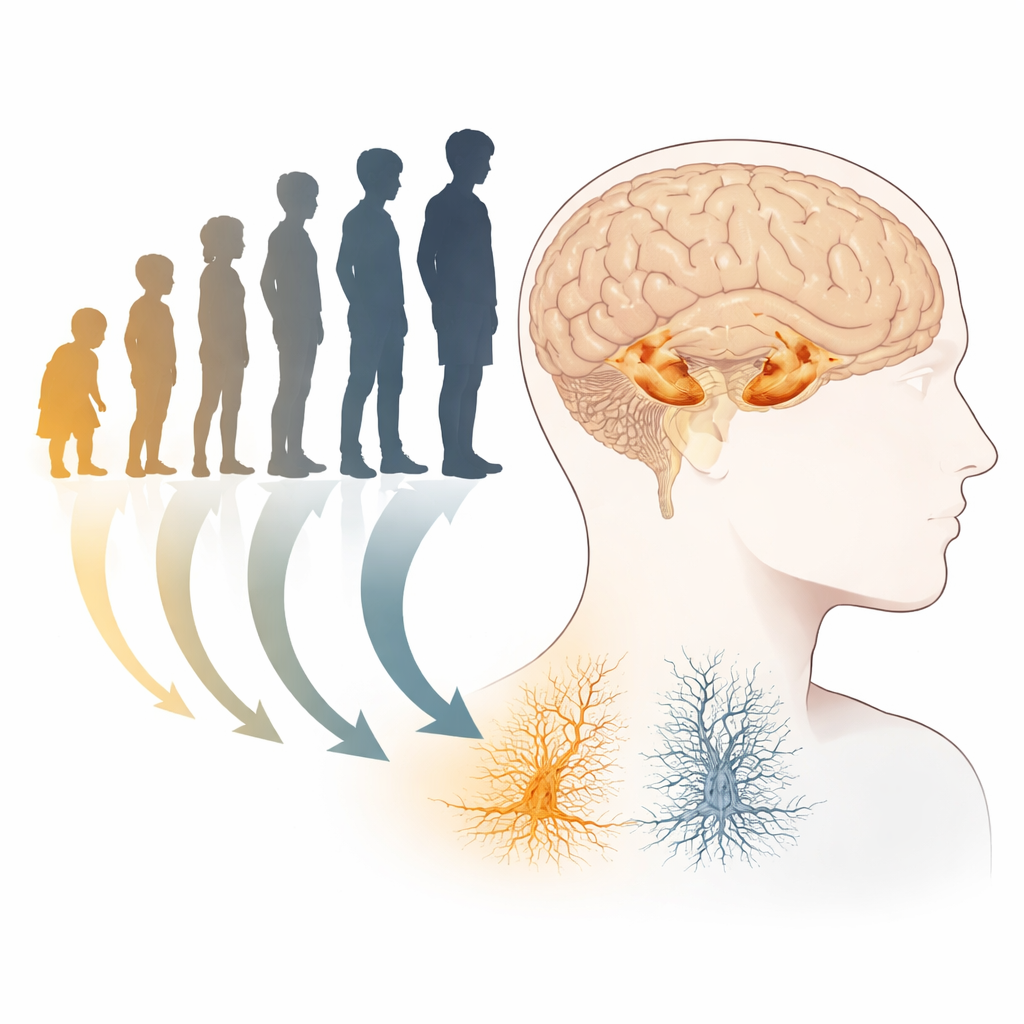
The hippocampus is a small, curved structure buried deep in the brain that helps us form memories, navigate spaces, and manage emotions. Childhood and adolescence are years of dramatic mental growth, yet scientists still know surprisingly little about how the fine wiring inside the hippocampus changes during this period. This study uses a powerful type of MRI to look beneath the surface, asking not just whether the hippocampus gets bigger, but how its inner circuits mature between ages 8 and 19.
Looking under the brain’s surface
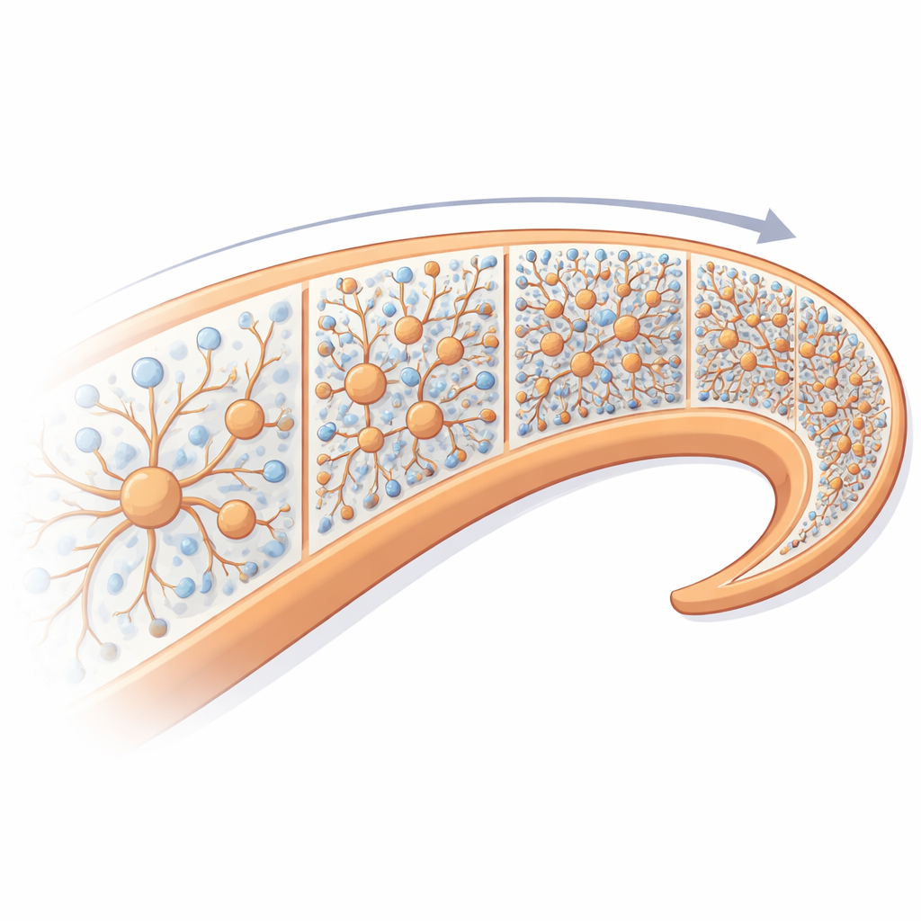
Most earlier research has treated the hippocampus like a single lump of tissue and focused on its overall size. Those studies have produced mixed results about whether it grows, shrinks, or stays steady in late childhood and adolescence. In this work, the researchers went beyond simple volume. They scanned 88 healthy children and teens with an MRI scanner equipped with ultra-strong magnetic gradients, which makes it possible to track the tiny movements of water molecules inside brain tissue. By analyzing how water diffuses, they could infer features of the microscopic wiring: the long, thin branches of nerve cells (neurites), their cell bodies (somas), and the spaces between them.
Peering into the hippocampal maze
The team used a specialized tool to "unfold" the hippocampus into a smooth surface, letting them map measurements across its different subregions and along its front-to-back axis. They applied several advanced models of diffusion, including one called SANDI, which estimates how much of the MRI signal comes from neurites, from somas, and from the surrounding fluid-filled space. In parallel, they measured more familiar diffusion markers like mean diffusivity, which captures how freely water moves. This combination allowed them to test whether age-related changes show up mainly in gross shape (thickness, folding, and volume) or in the underlying microstructure.
Hidden growth without visible expansion
Despite the age span from late childhood into late adolescence, the overall size, thickness, and surface folding of hippocampal subregions barely changed. In contrast, the microstructural markers shifted robustly with age. Across nearly all subfields and along most of the long axis, the fraction of signal attributed to neurites increased, while the fraction linked to extracellular space and the average apparent soma radius decreased. Water diffusion became more restricted, consistent with a denser, more complex internal network of branches. These trends suggest that even when the hippocampus no longer grows outward, it is still being remodeled inside, with tighter packing of neural processes and possibly more myelin and synapses.
Differences along the structure and between sexes
The study also found that not all parts of the hippocampus mature in the same way. Some microstructural changes varied more between its classic subfields, while others lined up more clearly along the front-to-back (anterior–posterior) axis. Orientation analyses showed that the preferred direction of water diffusion shifted with age in specific regions, hinting at reorganization of internal pathways. When the researchers compared boys and girls, they observed different age-related trends for several measures: in general, male participants showed more pronounced increases in certain structural features, whereas changes in females appeared earlier and then leveled off. These differences may reflect the influence of puberty and sex hormones on brain development.
Linking MRI signals to real cells
To interpret what these MRI-based changes might mean biologically, the authors compared their age-related maps with high-resolution data from adult human brain tissue, including staining for myelin, nerve fibers, various types of inhibitory neurons, and a marker of synapse density measured with PET imaging. Regions where neurite-related signal increased most with age tended to be areas that, in adults, have more myelin and more synapses. Conversely, places where water remained more free to diffuse were associated with lower myelin content. These patterns support the idea that the diffusion changes seen in children and adolescents are tracking real, long-lasting refinements in wiring, insulation, and connectivity.
What this means for growing minds
For a layperson, the key message is that during late childhood and adolescence, the hippocampus is not simply getting bigger; it is getting more intricate. Even as its outer shape stays largely stable, the internal forest of branches grows denser and more refined, especially in ways linked to myelin and synapses. These micro-level changes likely support the steady improvement in memory, thinking, and emotional regulation that marks the journey from childhood to adulthood. Understanding this hidden remodeling may eventually help scientists identify when development goes off track and could inform earlier interventions for learning and mental health conditions that involve the hippocampus.
Citation: Karat, B.G., Genc, S., Raven, E.P. et al. Microstructural variation of hippocampal substructures across childhood and adolescence quantified with high-gradient diffusion MRI. Commun Biol 9, 416 (2026). https://doi.org/10.1038/s42003-026-09622-x
Keywords: hippocampus development, adolescent brain, diffusion MRI, brain microstructure, memory and cognition