Clear Sky Science · en
Characterization of mouse melanocytes reveals ultrastructural and immunological insights into the inner ear function
Why tiny pigment cells in the ear matter
Deep inside the inner ear, tiny pigment‑producing cells called melanocytes sit next to sound‑sensing and balance‑sensing structures. Doctors have long noticed that people and animals with pigment disorders often have hearing or balance problems, but the details have been murky. This study uses high‑resolution microscopy and modern genetic tools in mice to map out exactly what these pigmented cells are, how they change with age and injury, and how they might help protect hearing.
Sorting out look‑alike cells
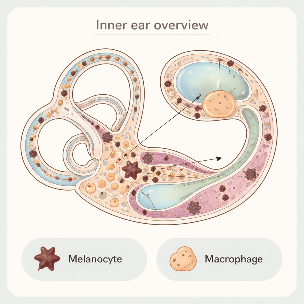
Under a regular microscope, several cell types in the inner ear look dark because they contain small pigment granules or other dense material. Earlier work proposed a strange hybrid cell in the cochlea’s stria vascularis—a crucial tissue for generating the ear’s electrical "battery"—called a perivascular macrophage‑like melanocyte. The new study shows that this hybrid does not actually exist. Using special stains and electron microscopes, the authors separate true melanocytes from nearby immune cells called macrophages. Melanocytes have an "octopus‑like" shape with long extensions and scattered pigment packages, and they carry classic melanocyte markers. Macrophages are rounder, sit close to blood vessels, and show immune markers; when they contain pigment, it is because they have swallowed melanin granules rather than made them.
Pigment and cleanup crews in the balance organs
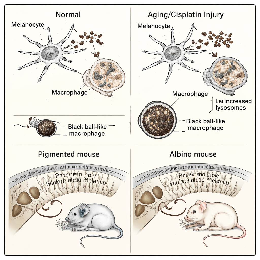
In the vestibular part of the inner ear, which helps control balance, the team found another surprise. Large, very dark, round "black ball" structures had often been assumed to be pigment cells. Closer inspection revealed that these too are macrophages stuffed with pigment they have engulfed. True vestibular melanocytes lie nearer to the supporting membrane, are flatter, and send processes across the membrane toward the fluid‑filled space, suggesting they may release melanin or other molecules into the inner‑ear fluids. Over time, especially as mice age, the number of melanocytes in these regions falls, while the number of "black ball" macrophages rises, implying that macrophages are clearing away dying melanocytes and their pigment.
Changes with age, drugs, and coat color
The researchers then asked how these cells behave under stress. With aging and after treatment with the chemotherapy drug cisplatin, macrophages in the cochlea and vestibule become more numerous and more active, and they contain many more pigment granules and lysosomes—the cell’s recycling centers. Meanwhile, melanocytes lose pigment and show signs of wear. Comparing dark‑furred mice to albino mice, which make little melanin, revealed that albino animals have fewer fully matured pigment granules and their macrophages pick up less melanin overall. Functionally, pigmented mice recovered their hearing better after loud noise exposure than albino mice, supporting the idea that melanin helps buffer damaging changes—such as surges in ions like potassium and calcium or binding of toxic drugs—and so protects inner‑ear cells.
Tracing pigment pathways in a mutant mouse
To explore how melanocytes get to their final positions during development, the team studied a mutant strain lacking a key gene, Pou3f4, which shapes the ear’s supporting tissue. These mice showed odd pigment patterns: excess melanin in the central bony core and balance organs, but fewer melanocytes and a thinner pigment layer in the stria vascularis, along with early‑aging‑like changes in macrophages. From where pigment accumulated, the authors propose that melanocytes normally migrate from the central core of the cochlea along a structure called Reissner’s membrane and then spread from the base toward the apex. When this journey is disrupted, fewer melanocytes reach the stria vascularis, potentially weakening the ear’s power supply for hearing.
What this means for hearing health
Seen together, the results redraw the cellular map of pigment and immune cells in the inner ear. Rather than exotic hybrid cells, there are two cooperating but distinct players: melanocytes that make melanin and help maintain the ear’s delicate chemical balance, and macrophages that act as janitors, engulfing excess or damaged pigment and cellular debris. With age, genetic changes, or exposure to toxic drugs and loud noise, this partnership shifts: melanocytes dwindle and macrophages become heavily loaded with pigment. For non‑specialists, the key message is that these tiny pigment cells are not cosmetic extras—they are working parts of the machinery that keeps hearing and balance stable, and understanding them better may eventually guide new strategies to protect the inner ear from damage.
Citation: Cai, J., Xu, L., Song, Y. et al. Characterization of mouse melanocytes reveals ultrastructural and immunological insights into the inner ear function. Commun Biol 9, 325 (2026). https://doi.org/10.1038/s42003-026-09616-9
Keywords: inner ear, melanocytes, macrophages, hearing loss, melanin