Clear Sky Science · en
Neural mechanisms of feature binding in working memory
How the Brain Keeps Our Experiences Together
When you remember a scene—say, a red mug on the right side of your desk—you don’t just store “red” and “mug” and “right” separately. Your mind somehow glues these pieces into a single, vivid memory. This paper asks a deceptively simple question: how does the brain actually pull off that glue work, known as feature binding, in our short-term or “working” memory? Understanding this process can shed light on everyday abilities like recognizing objects, following directions, and perhaps why memory sometimes fails with age or disease.
From Separate Bits to Unified Moments
Our visual world is made of separate features—colors, shapes, and locations—that must be combined so we can recognize objects and remember what went where. Classic theories propose that attention helps link features onto a shared spatial map. Yet earlier brain imaging studies pointed to many different regions—hippocampus, frontal and parietal areas, even early visual cortex—without clearly explaining how they work together. A major problem was that past experiments often compared memories of combined features against memories of just one feature, unintentionally changing how much information people had to remember.
A Fair Test of Memory Glue
To fix this, the researchers scanned the brains of 40 volunteers while they performed a picture-based memory game. On each trial, people briefly saw several colored disks at different locations, then had to hold both color and position in mind during a pause. In one condition, they needed to remember the exact color–location pairings (true bindings). In another, they still remembered both color and location, but only had to answer about one or the other at test, so the features could be kept separate. This clever design kept the total amount of information the same in both conditions, isolating the extra mental work of gluing features together.
More Brain Teamwork, Not Just More Activity
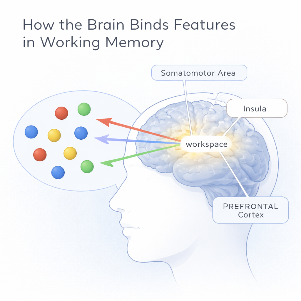
The team used functional MRI to track where blood flow—and thus brain activity—increased. Surprisingly, when they compared the two conditions directly, no single region lit up significantly more for bindings than for separate features. Instead, both tasks activated a broad set of areas, including prefrontal cortex, regions near the central sulcus (involved in movement and sensation), the insula, and parietal–temporal visual areas. To dig deeper, the researchers treated the brain as a network, using graph theory to ask how efficiently different regions exchanged information. During binding, eight areas showed higher “local efficiency,” meaning they were better at relaying and processing information within their immediate neighborhood. These key sites included extrastriate visual cortex, the somatomotor area, inferior parietal lobe, both insulas, and several parts of prefrontal cortex and retrosplenial cortex.
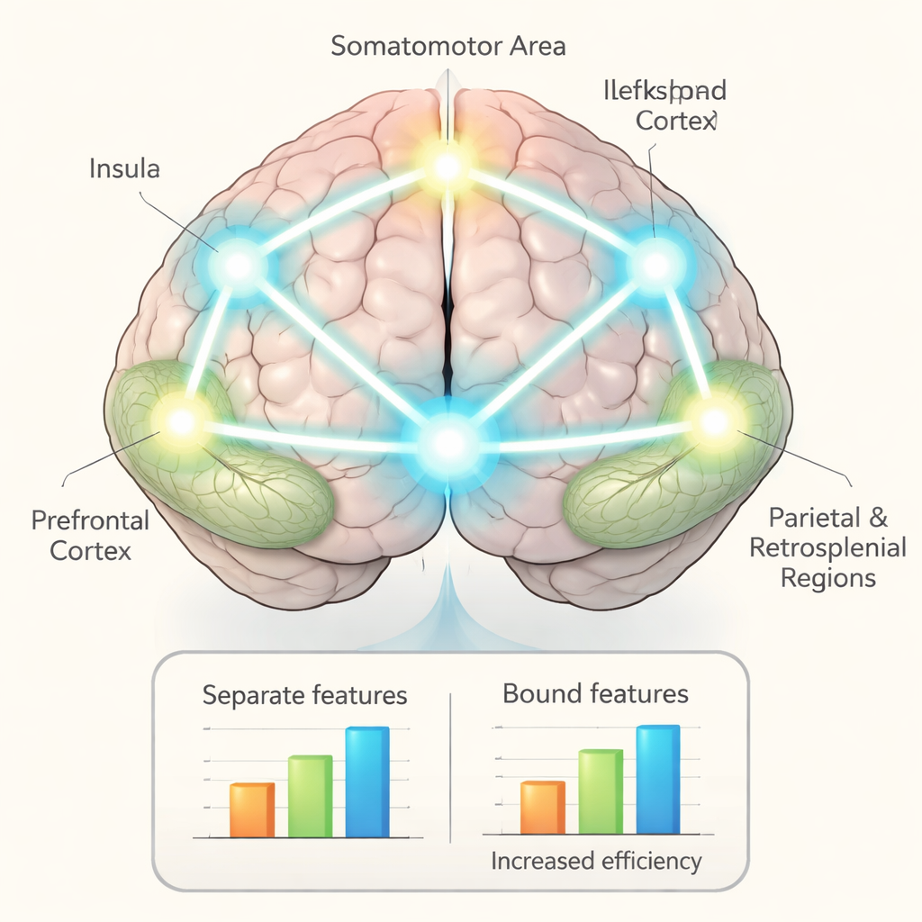
A Central Workspace with a Fast Starter
Focusing on this eight-region set, the authors mapped how strongly each area was functionally connected to the others. They found a tightly linked “workspace” in which seven of the regions formed a cluster with stronger connections when people bound features together than when they kept features separate. The somatomotor area, prefrontal cortex, and insulae emerged as hubs, with many of the strongest links running through them. The somatomotor area stood out in another way: its activity fluctuated over the shortest timescale, suggesting it responds quickly to incoming visual information, then passes signals on to slower, more stable regions like the insula and prefrontal cortex. Stronger connections from the somatomotor area to these regions were also tied to longer reaction times, consistent with the idea that binding requires extra processing steps.
Why This Matters for Everyday Memory
Put simply, the study suggests that remembering “what was where” is not handled by a single memory center, but by a cooperative network acting as a central workspace. In this workspace, the somatomotor area seems to kick off fast, early processing, while the insula and prefrontal cortex help stabilize and maintain the bound representations over time. This extra coordination makes binding slightly slower and more demanding than remembering features in isolation, but it is also what allows us to hold together the rich, detailed scenes of everyday life. Understanding this network may eventually help explain why feature binding breaks down in some neurological conditions, and could guide new approaches to support or restore everyday memory.
Citation: Cao, Y., Chen, F., Wang, H. et al. Neural mechanisms of feature binding in working memory. Commun Biol 9, 270 (2026). https://doi.org/10.1038/s42003-026-09548-4
Keywords: working memory, feature binding, brain networks, attention, visual perception