Clear Sky Science · en
Widefield cortical activity and functional connectivity during motorized locomotion
How Walking Shapes the Brain
Every step we take relies on a constant conversation between our senses and our muscles. But scientists still don’t fully understand how the brain keeps us moving smoothly when the ground beneath us changes. This study peered across the outer surface of the mouse brain while the animals walked on different kinds of moving tracks, revealing that the brain’s communication patterns—not just its overall activity—shift depending on how the body must move.
Three Ways to Take a Walk
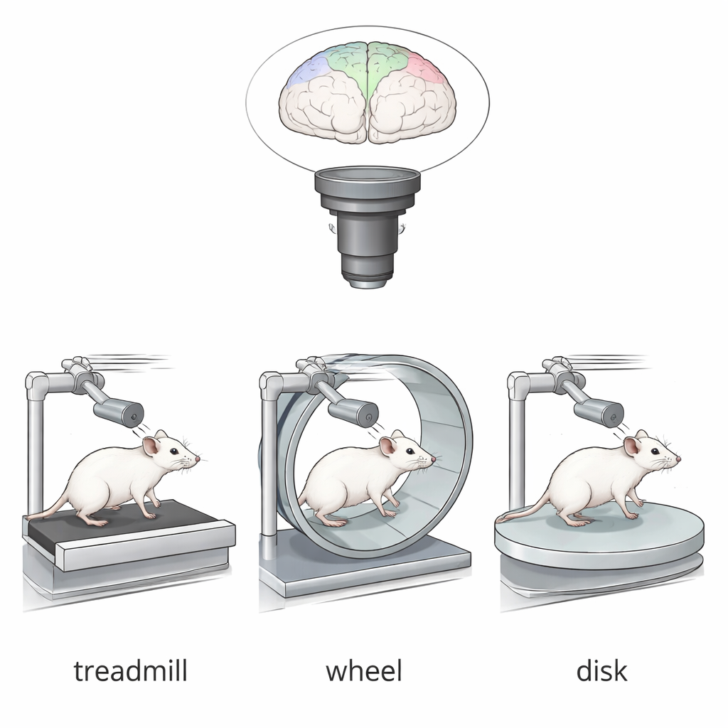
To explore how the walking environment affects the brain, the researchers trained mice to walk while their heads were gently fixed in place. The animals walked on three motorized tracks: a flat treadmill belt, a curved running wheel, and a rotating disk that spun around a central point. All three required the mice to keep up with a moving surface, but each demanded different stepping patterns and balance. While the mice walked, a transparent “window” in the skull allowed the team to use widefield calcium imaging—a method that makes active nerve cells glow—to monitor activity across nearly the entire top surface of the brain in real time.
Separating Movement from Inner Commands
Raw brain signals during walking are a mix of two things: the brain’s own internal motor commands and the sensory and body signals generated by moving limbs, changing posture, and shifting arousal. To untangle these, the researchers tracked the animals’ hindlimb joints and pupil size using high-speed cameras and modern pose-tracking software. They then used a statistical method called partial least squares regression to mathematically remove the influence of these measured body variables from the brain activity. The remaining signal—what they call “internally driven” activity—reflects how the brain organizes movement from within, beyond the direct echoes of limb motion and eye dilation.
Same Overall Activity, Different Conversation Patterns
One surprising finding was that the average level of internal activity across major brain areas during steady walking was quite similar, no matter which track the mice used. Regions involved in movement and sensation, such as primary and secondary motor cortex and somatosensory cortex, all became active when walking began and quieted when it ended. However, when the team looked at how these regions co-fluctuated—that is, how strongly their activity rose and fell together—the story changed. The pattern of “functional connectivity” across the cortex depended strongly on the track type, even though the overall activity levels did not.
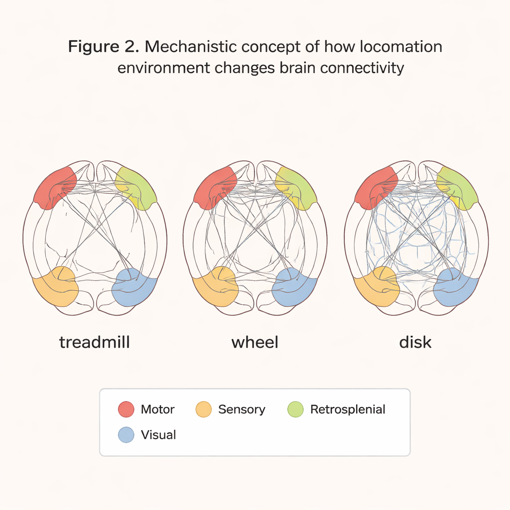
A Special Role for a Motor Planning Hub
The secondary motor cortex, or M2, is thought to help transform sensory information into plans for movement. During sustained walking on the treadmill, this medial part of M2 showed distinctly weaker internal connectivity with the rest of the cortex compared with walking on the wheel or disk. On the curved wheel and rotating disk, where the animals had to constantly adjust posture and trajectory, M2 and distant regions like the visual and retrosplenial cortices were more tightly linked. On the simpler, straight treadmill, by contrast, M2’s reduced coupling suggests that, once a stable gait is reached, it may shift into an inhibitory or gating role, limiting unnecessary communication while the body executes a well-practiced pattern.
Why the Shape of the Ground Matters
Overall, the study shows that the brain’s internal communication network during walking is tuned to the physical demands of the environment. Linear tracks like treadmills produce relatively stable locomotion with reduced need for complex coordination, while curved or rotating tracks drive richer interactions between motor, sensory, and navigation-related regions. For researchers and clinicians interested in movement disorders or rehabilitation, this work highlights that not all walking tasks are equal: understanding health and disease will require paying attention not only to how active the brain is, but also to how its regions talk to one another under different kinds of movement challenges.
Citation: Lee, C.H., Lee, G., Song, H. et al. Widefield cortical activity and functional connectivity during motorized locomotion. Commun Biol 9, 264 (2026). https://doi.org/10.1038/s42003-026-09541-x
Keywords: locomotion, motor cortex, functional connectivity, sensory-motor integration, widefield imaging