Clear Sky Science · en
How do infant brains fold? Sulcal deepening is linked to development of sulcal span, thickness, curvature, and microstructure
How Baby Brains Get Their Folds
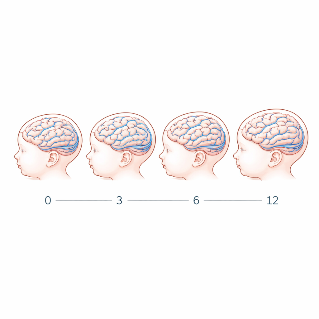
From the moment a baby is born, their brain is rapidly reshaping itself. One of the most striking changes is how the smooth-looking surface becomes covered with deeper and more complex folds. These folds are not just wrinkles: they help pack more brain tissue into the skull and are closely tied to how thinking, seeing, moving, and social skills emerge. This study asks a simple but fundamental question: during the first year of life, how exactly do those folds deepen, and what changes in brain tissue go along with that process?
The Hidden Landscape Inside an Infant’s Brain
Deep grooves in the brain, called sulci, separate the raised ridges known as gyri. Many sulci already appear before birth, but they continue to change dramatically after a baby is born. The researchers used safe, noninvasive MRI scans on 43 full-term infants, some scanned more than once between birth and 12 months, for a total of 79 sessions. They focused on 15 long, well-defined sulci spread across the brain—regions involved in vision, touch, movement, language, and higher thinking. By tracking these same grooves over time, they could measure how their shape and the underlying tissue changed during the first year.
Early Folds vs. Late Folds
Not all brain folds follow the same timetable. Some sulci form early in pregnancy, while others appear later. The team found a striking pattern: sulci that emerge earlier in the womb start out deeper at birth but change relatively little after. Later-appearing sulci, in contrast, are quite shallow in newborns but then deepen rapidly during the first year. Overall, average sulcal depth increased by about 21 percent, especially in the first six months. This suggests that the brain’s folding schedule is set early, but different sulci continue their “growth spurts” at different times after birth.
Growing Wider, Thicker, and Less Curved
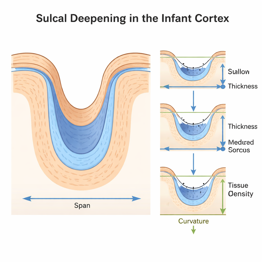
The study did more than measure depth. The scientists also looked at how wide each groove was (its span), how thick the surrounding cortex was, how sharply it curved, and how dense the tissue inside became. Across the first year, sulci widened by about 42 percent, and the cortex around them thickened by about 21 percent. At the same time, the folds became less tightly curved—more open and less pinched. Yet these changes did not simply mirror when each sulcus first appeared during pregnancy. Instead, sulci that were already thick at birth changed less, while thinner ones thickened more, following their own tempo.
Inside the Tissue: Building Denser Brain Matter
Using a special MRI measure called R1, which reflects how dense and myelin-rich brain tissue is, the researchers showed that the gray matter inside sulci becomes about one-third denser in the first year. This microstructural growth did not follow a single global pattern either: different sulci showed different rates of tissue change. When the scientists combined all measures—span, thickness, curvature, and tissue density—they found that depth could be predicted as a weighted mix of these factors, and that the exact recipe varied from one sulcus to another. In other words, there is no single “knob” that controls folding; instead, multiple physical and biological processes work together.
Zooming In: Why the Deepest Points Matter Most
Within each sulcus, the deepest line down the middle—the fundus—stood out. Local analyses showed that deeper points along a sulcus tend to have sharper curvature, denser tissue, and slightly thinner cortex than the surrounding walls. These subtle differences suggest that fundi may be special zones where tissue growth, mechanical forces, and future brain functions converge. Previous work has linked such deep regions to key abilities, like hand movement, language, and face or place recognition, raising the possibility that the physical shaping of these folds helps organize how mental functions are laid out.
What This Means for Early Brain Health
For a non-specialist, the main takeaway is that infant brain folding is both orderly and diverse. Some sulci mature early and change little; others keep reshaping themselves throughout the first year. Depth is closely tied to how wide folds are, how thick and curved the cortex is, and how densely packed its tissue becomes. By mapping these normal trajectories in detail, this work provides a reference chart for healthy brain development. Because unusual patterns of sulcal depth have been linked to conditions like autism, Down syndrome, and depression, such benchmarks may eventually help doctors spot early deviations and understand how the physical architecture of the infant brain supports the emergence of thought, perception, and behavior.
Citation: Tung, S.S., Yan, X., Fascendini, B. et al. How do infant brains fold? Sulcal deepening is linked to development of sulcal span, thickness, curvature, and microstructure. Commun Biol 9, 258 (2026). https://doi.org/10.1038/s42003-026-09536-8
Keywords: infant brain development, cortical folding, sulcal depth, brain MRI, neurodevelopmental disorders