Clear Sky Science · en
Closed loop text guided framework for lung cancer lesion segmentation and quantification
Why this matters for lung cancer care
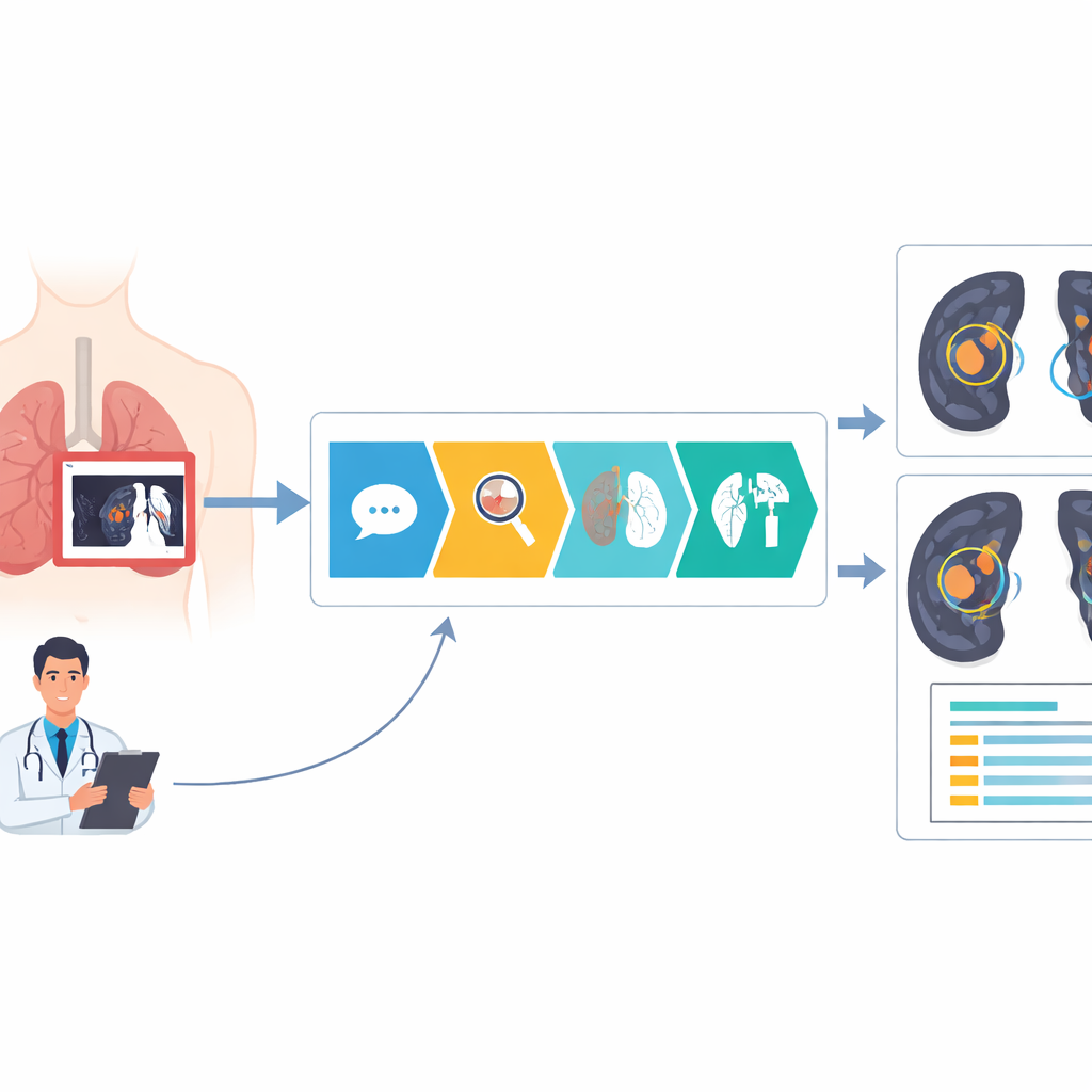
Lung cancer kills more people worldwide than any other cancer, in large part because tumors are often found late or measured imprecisely. Many hospitals, especially in low-resource settings, now have CT scanners, but lack enough experts to interpret every scan quickly and consistently. This study introduces BiomedLoop, an artificial intelligence system designed to read lung CT images in a way that speaks the same “language” as radiologists, aiming to find and measure lung tumors more accurately while producing report-ready information.
From words on a screen to spots in the lung
Radiologists typically describe what they see in free-form text: for example, a small nodule in the upper right lung. Traditional computer methods instead work only with raw pixels, drawing mask-like outlines that don’t easily connect back to everyday medical descriptions. BiomedLoop bridges this gap. It takes short text phrases similar to those in reports and uses a localization module to find likely regions on the CT scan where the described lesion might be. A second module then refines these coarse regions into detailed shapes that follow the true tumor boundaries, bringing computers a step closer to how human experts think about “where” a lesion is.
Turning outlines into numbers and back into meaning
Once BiomedLoop has traced a tumor, it does more than just shade in the abnormal area. The system converts each outline into concrete measurements such as the tumor’s volume, what fraction of the lung it occupies, and its 3D position inside the chest. These measurements are then turned into structured, report-style text templates that mimic how radiologists summarize findings. Crucially, the system feeds these auto-generated descriptions back into its own learning process. By repeatedly pairing its measured outlines with their matching phrases, BiomedLoop improves its ability to connect language, images, and geometry—even on datasets that never had written reports in the first place.
Sharpening blurry edges with uncertainty
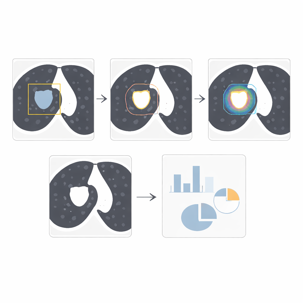
One of the hardest parts of lung cancer imaging is deciding exactly where a tumor ends and normal lung tissue or blood vessels begin, especially when the borders are faint. BiomedLoop introduces a special feature that focuses extra attention on these uncertain boundary regions. Instead of treating every pixel equally, the model first estimates where it is most unsure and allocates more computing power there, leaving less ambiguous areas to a lighter process. This uncertainty-aware strategy stabilizes the outlines, cutting down on jagged or misplaced borders that can distort size estimates. The end result is cleaner, more consistent tumor contours, even when the images are noisy or the lesions are subtle.
Proving its strength across many datasets
The researchers tested BiomedLoop on five independent public lung cancer datasets, comparing it with widely used neural networks and with newer “segment anything” models adapted from general computer vision. They measured how much the computer-drawn tumor shapes overlapped with expert annotations and how close the boundaries were in millimeters. Across most datasets, BiomedLoop achieved the highest overlap and the smallest boundary errors, while also aligning better with the initial text or box prompts used to guide it. Importantly, these gains held up when the system was moved from one hospital’s data to another’s, suggesting that it can generalize well across different scanners, imaging protocols, and patient populations.
What this means for patients and clinicians
For non-specialists, the key takeaway is that BiomedLoop can turn a radiologist-like description into a precise tumor outline and back into standardized, machine-readable measurements. This closed loop—from text to image to numbers and again to text—could reduce the need for tedious manual contouring, support more consistent treatment planning, and allow large numbers of scans to be processed quickly in settings with few experts. While real-world trials are still needed, the study shows that combining language and imaging in a single system can make AI tools more explainable and clinically usable, offering a promising path toward faster, fairer lung cancer diagnosis and follow-up everywhere.
Citation: Wang, S., Wang, Z., Men, W. et al. Closed loop text guided framework for lung cancer lesion segmentation and quantification. npj Digit. Med. 9, 237 (2026). https://doi.org/10.1038/s41746-026-02422-x
Keywords: lung cancer imaging, medical AI, text-guided segmentation, CT scan analysis, tumor quantification