Clear Sky Science · en
Neck-to-knee dixon MRI thigh volume as a superior mass biomarker for Sarcopenia: evidence from the UK biobank
Why leg muscles matter as we age
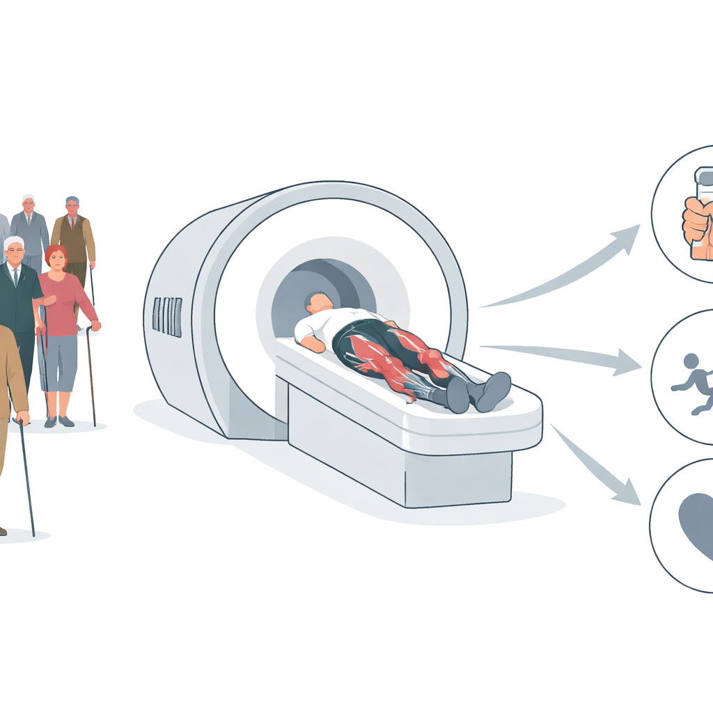
As people grow older, many worry about losing strength, balance, and independence. A key player in this story is sarcopenia, the gradual loss of muscle that raises the risk of falls, fractures, and even early death. This study asks a simple but important question: are we looking at the right muscles, in the right way, when we judge who is at risk? By using advanced MRI scans and artificial intelligence on tens of thousands of adults, the researchers show that the detailed shape and balance of the thigh muscles—not just how much muscle we have overall—can better flag who is likely to become weak or frail.
Looking beyond one thin slice
Today, doctors and researchers usually estimate muscle mass with whole-body X-ray scans (DEXA) or a single “slice” of a CT scan taken through the abdomen. These methods give a rough total of lean tissue but blur together many different muscles and often focus on the trunk rather than the legs. That is a problem, because everyday movements—standing up from a chair, climbing stairs, catching yourself from a stumble—depend heavily on the large muscles of the thighs. A single cross-section near the spine cannot capture how those leg muscles are arranged along their length, nor can it separate the muscles that straighten the knee from those that bend the hip. The authors argue that to understand real-world mobility, measurements must follow the actual working machinery: the muscles that move our legs.
Turning whole-leg scans into usable numbers
Manually tracing every muscle in hundreds of MRI images for each person would be impossibly slow. To overcome this, the team built an automated system based on a modern “transformer” neural network that can recognize and outline 27 different muscles and bones from the pelvis to the knee. They applied this system to neck-to-knee MRI scans from 37,004 participants in the UK Biobank, a long-running health study of middle-aged and older adults. The computer’s segmentations closely matched expert labels and showed excellent agreement with standard DEXA measures of leg lean mass, confirming that the new method produces reliable volume estimates for the thigh muscles as a whole.
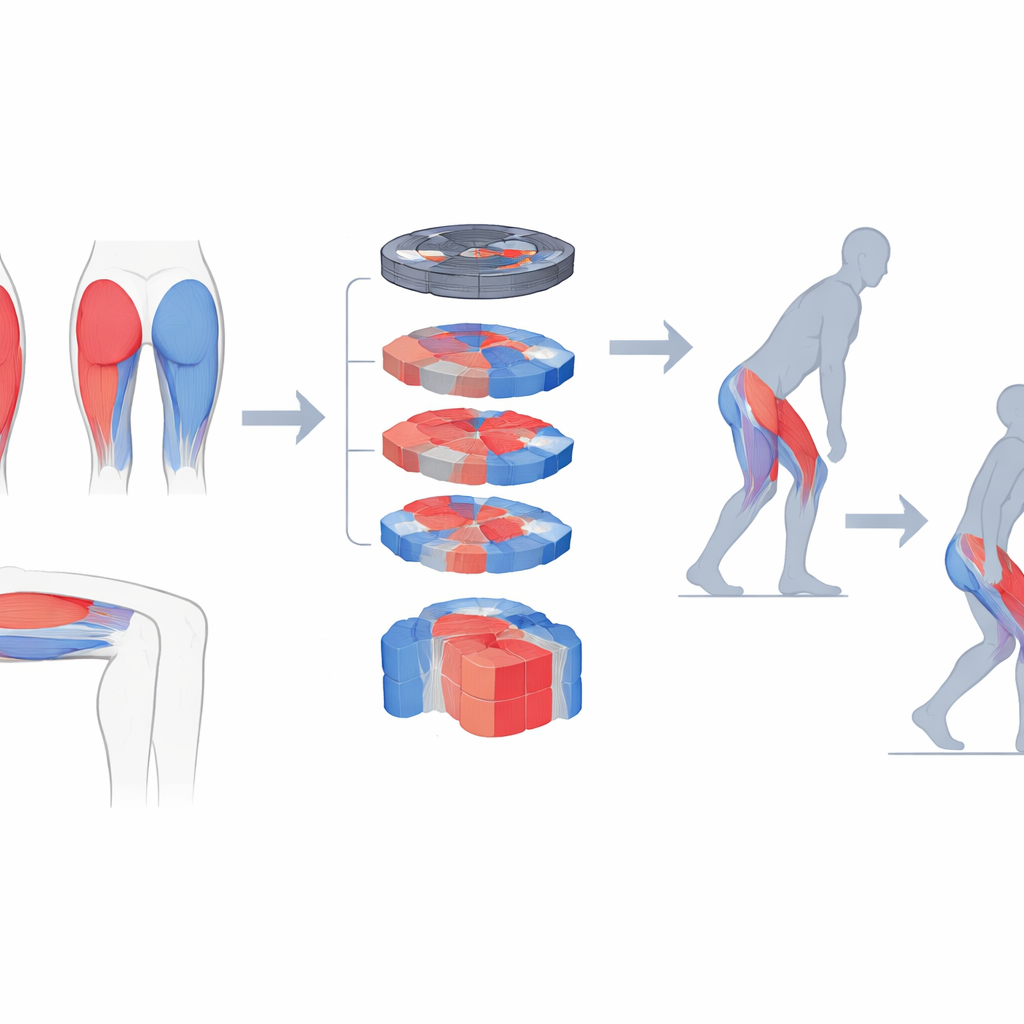
Front-versus-back balance as a warning sign
Having validated their tool, the researchers moved beyond simple muscle totals to examine how mass is distributed within the thigh. They compared the bulk of the muscles on the front of the thigh, which straighten the knee (the quadriceps), with those on the back, which help extend the hip (the hamstrings and gluteals). This produced a simple front–back balance measure. People whose thighs were relatively “back heavy”—with less muscle volume in the front compared to the back—were much more likely to have weak handgrip, be classified as sarcopenic, report recent falls, and to have died during follow-up. These links remained even after accounting for age, sex, body size, and activity levels, and they appeared despite having similar overall muscle mass. In contrast, left–right differences between legs showed little relationship to weakness or falls, suggesting that front–back balance matters more than side-to-side symmetry.
A new, richer picture of muscle aging
The study also tracked how various muscle measures changed with age in men and women. As expected, total muscle volume and standard DEXA indices declined steadily over the decades. However, the MRI-based measures revealed growing variation in later life: while some older adults maintained relatively robust thigh muscles, others showed sharp losses or imbalanced patterns. This spread hints at different “aging trajectories,” where individuals with similar weights or total lean mass may follow very different paths in muscle health. Because the same MRI scans can also be used to map fat content inside muscles, the authors argue that future work could combine quantity, quality, and distribution into a single, richer description of muscle status for each person.
What this means for healthy aging
For non-specialists, the main message is that where muscle sits in the thigh can be as important as how much muscle there is. Losing more of the front-of-thigh muscles that help you rise, climb, and catch yourself may quietly raise the risk of weakness, falls, and premature death, even if overall muscle mass seems acceptable. By pairing large-scale MRI scans with automated analysis, this work offers a practical way to spot high-risk muscle patterns in big populations and, eventually, in clinics. In the long term, such detailed “muscle maps” could guide more precise exercise and rehabilitation programs—aimed, for example, at rebuilding vulnerable front-of-thigh muscles—so that older adults can stay steady, strong, and independent for longer.
Citation: Kim, H.S., Park, H., Kang, J. et al. Neck-to-knee dixon MRI thigh volume as a superior mass biomarker for Sarcopenia: evidence from the UK biobank. npj Digit. Med. 9, 239 (2026). https://doi.org/10.1038/s41746-026-02379-x
Keywords: sarcopenia, thigh muscle volume, MRI segmentation, muscle distribution, falls in older adults