Clear Sky Science · en
Prompt-mamba filtering networks for accurate hepatocellular carcinoma lesion segmentation in abdominal CT
Why better liver scans matter
Liver cancer is one of the world’s deadliest cancers, in part because many tumors are hard to see clearly on routine medical scans. Radiologists use CT images to sketch the exact outline of each tumor, a painstaking task that directly shapes surgery, ablation, and follow-up care. This article presents a new artificial intelligence (AI) system, called Prompt-Mamba-AF, designed to automatically trace liver tumors more accurately and consistently than current tools, especially the tiny early-stage spots that are easiest to miss.
A new way to teach computers where to look
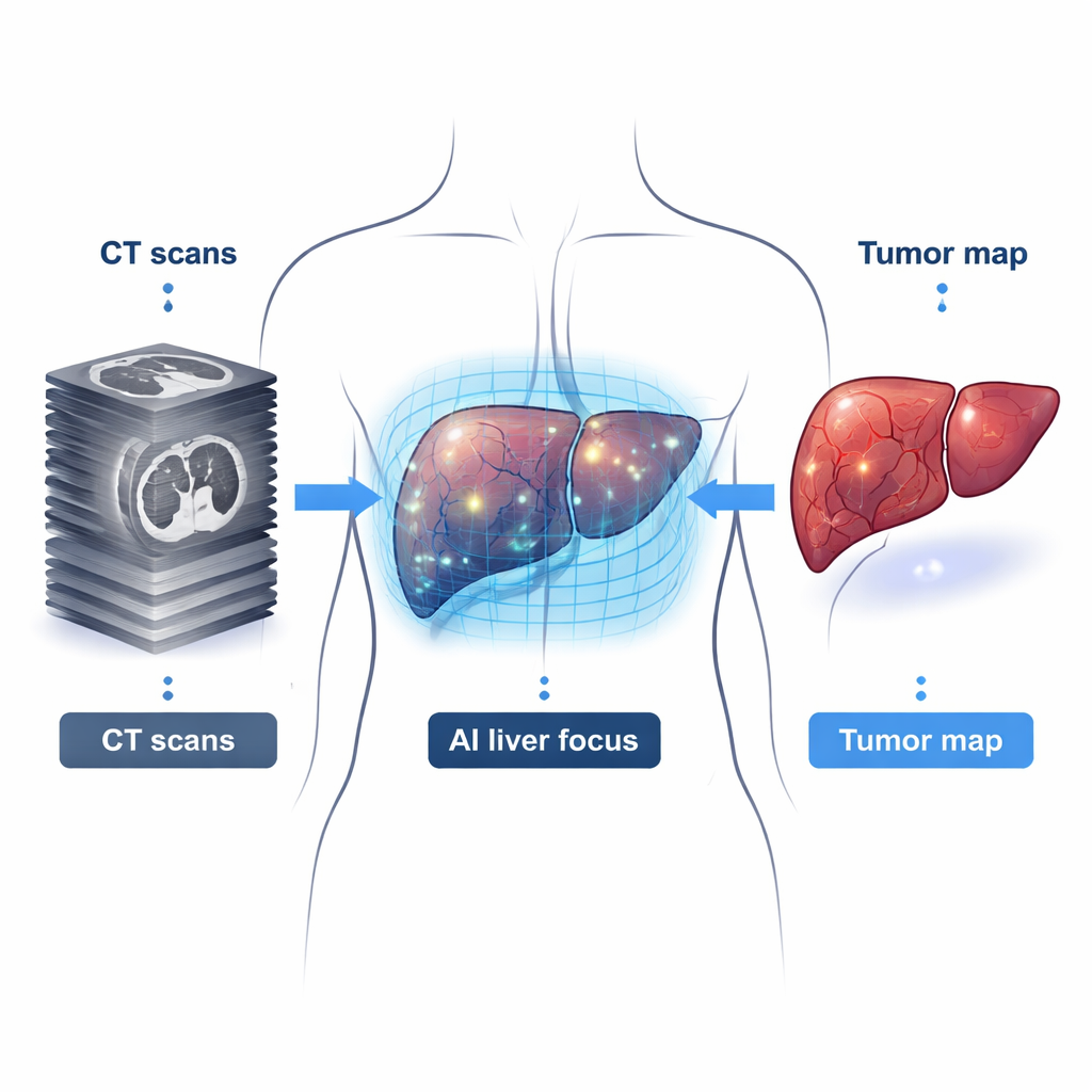
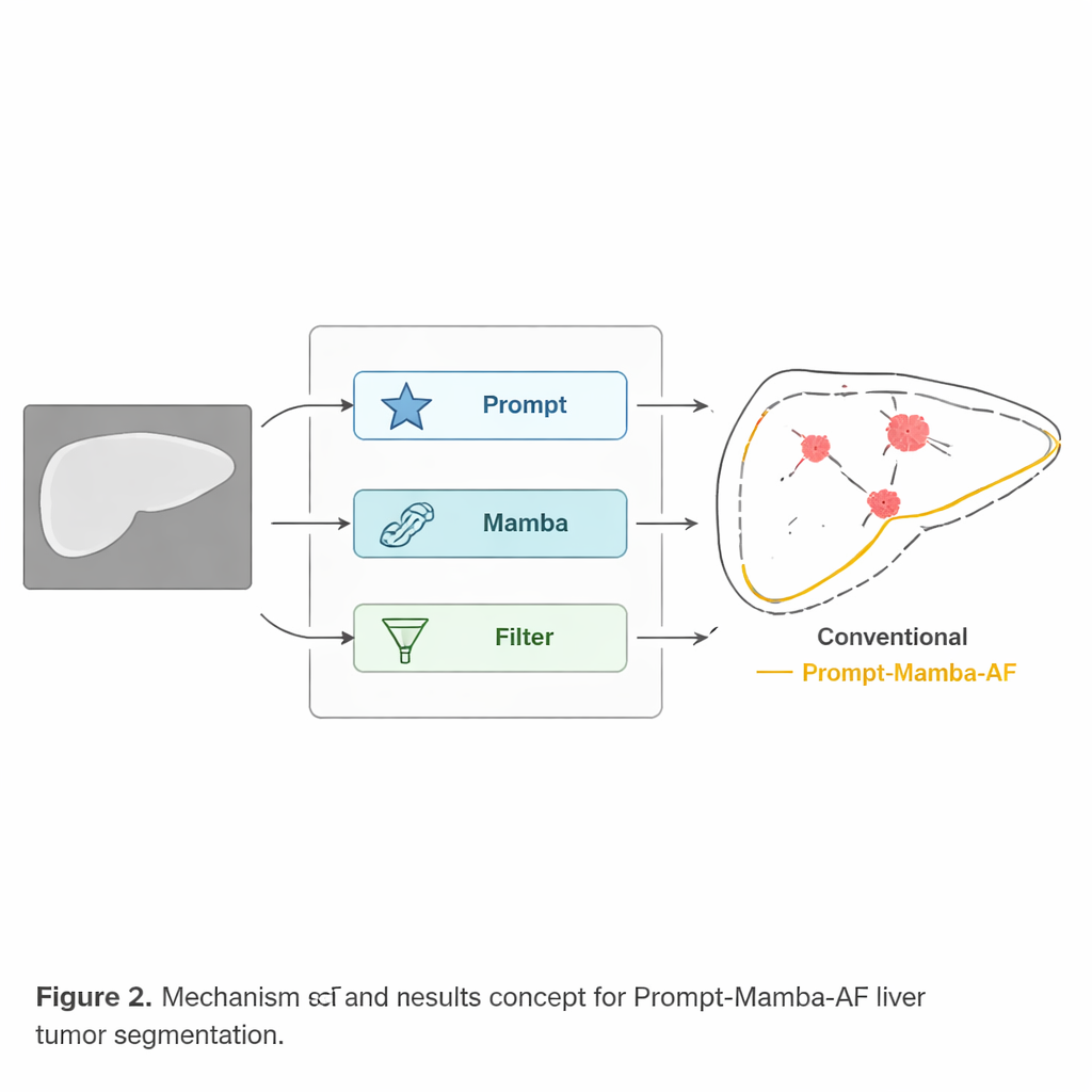
One core challenge in liver imaging is that tumors can be small, oddly shaped, and nearly the same shade as the surrounding tissue. Traditional AI systems try to learn everything directly from the raw image, which means they often waste effort examining the entire abdomen instead of focusing on the liver. Prompt-Mamba-AF adds an extra hint: a coarse mask showing where the liver is. This “prompt” guides the algorithm to pay most attention to the organ of interest, filtering out distracting background structures such as ribs, spleen, and intestine. By narrowing the search area before deeper processing begins, the system can dedicate more of its capacity to telling tumor from healthy liver.
Following subtle patterns across 3D volumes
CT scans are three-dimensional, made of many thin slices stacked together. A small cancerous nodule may only appear clearly when these slices are considered as a whole. Many existing neural networks either look at only a few slices at a time or rely on a heavy mathematical operation called self-attention, which becomes very slow and memory-hungry for full 3D volumes. Prompt-Mamba-AF instead uses a newer kind of sequence model, known as a state-space model, to link information across the entire scan with far less computation. This “Mamba” module efficiently tracks long-range structure, helping the system notice faint but consistent abnormalities and keep tumor boundaries smooth and continuous from slice to slice.
Sharper outlines, fewer misses, across many hospitals
The researchers tested Prompt-Mamba-AF on multiple public datasets collected at different hospitals and with different scanners. On a large international CT collection of liver tumors, the new method edged out a range of popular convolutional and Transformer-based networks on standard accuracy measures, while using fewer parameters than many of its competitors. It was especially strong at finding small tumors: in lesions under 5 cubic centimeters, it achieved the highest overlap with expert annotations and recovered more tiny nodules that other systems missed. When trained on one CT dataset and evaluated “as is” on a separate CT set, as well as on MRI scans, the model still performed best, suggesting it had learned general liver and tumor shapes rather than overfitting to one machine or site.
Built-in safeguards for messy, real-world images
Hospital scans are rarely perfect: noise from low-dose imaging, slight patient motion, and streaks from metal implants can all obscure details. To mimic these conditions, the team deliberately corrupted test images with synthetic noise, blur, and missing regions. All algorithms got worse, but Prompt-Mamba-AF degraded the least. The liver prompt helped the model ignore irrelevant artifacts outside the organ, while the Mamba module’s global view allowed it to infer tumor continuity even when parts of the outline were damaged. A separate structure-aware filtering step in the decoder further cleaned up jagged or fragmented edges, producing tumor contours that looked more like what a radiologist would draw.
Toward flexible, reusable medical AI
Beyond liver cancer, the authors explored how well their design transfers to other organs and imaging types without retraining. Using simple masks to indicate kidneys, heart chambers, or pancreas, the same network achieved strong performance on these new tasks, rivaling or surpassing models tailored for each organ. This suggests that separating “where to look” (the prompt) from “how to draw the boundary” (the core network) may be a powerful recipe for building general-purpose medical image tools.
What this means for patients
For non-specialists, the main message is that Prompt-Mamba-AF makes computer assistance in liver cancer care both more accurate and more practical. By focusing on the liver, efficiently reading entire 3D scans, and enforcing clean, realistic tumor outlines, it detects more small lesions and produces measurements that are more trustworthy across hospitals and scanners. In the long run, such systems could help radiologists catch liver cancer earlier, plan surgeries with greater confidence, and monitor treatment response more objectively, without requiring enormous computing resources or massive, generic “foundation” models.
Citation: Xia, L., Chen, HY., Cao, YW. et al. Prompt-mamba filtering networks for accurate hepatocellular carcinoma lesion segmentation in abdominal CT. npj Digit. Med. 9, 193 (2026). https://doi.org/10.1038/s41746-026-02371-5
Keywords: liver cancer, CT imaging, medical AI, tumor segmentation, hepatocellular carcinoma