Clear Sky Science · en
Noninvasive prediction of occult pT3a upstaging in localized ccRCC with radiogenomic insights and prognostic relevance
Why this matters for people with kidney tumors
When doctors find a kidney tumor, they must choose between removing just the tumor and nearby tissue or taking out the entire kidney. That decision depends on how far the cancer has really spread. The trouble is that scans sometimes miss early, hidden invasion beyond the kidney, so a tumor that looks less serious before surgery can turn out to be more dangerous afterward. This study introduces a new artificial intelligence (AI) tool, called RENALNet, that uses routine CT scans to better spot these concealed high‑risk tumors, with the goal of guiding safer surgery and follow‑up care.
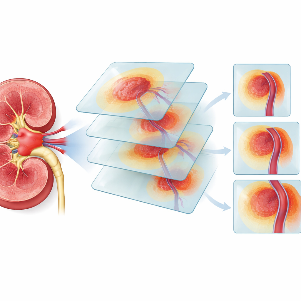
The hidden danger around the kidney
The researchers focused on clear cell renal cell carcinoma, the most common type of kidney cancer. Many of these tumors are found while they are still considered “localized,” meaning they appear to be confined to the kidney on imaging. Yet in 10–20% of such cases, detailed examination after surgery reveals that the cancer has already crept into the fat around the kidney or into nearby veins. This stage, called pT3a, is linked to a higher chance of the cancer coming back and of death. Standard CT or MRI scans often cannot see these tiny extensions, which means some patients may receive a kidney-sparing operation when a more extensive surgery would have been safer.
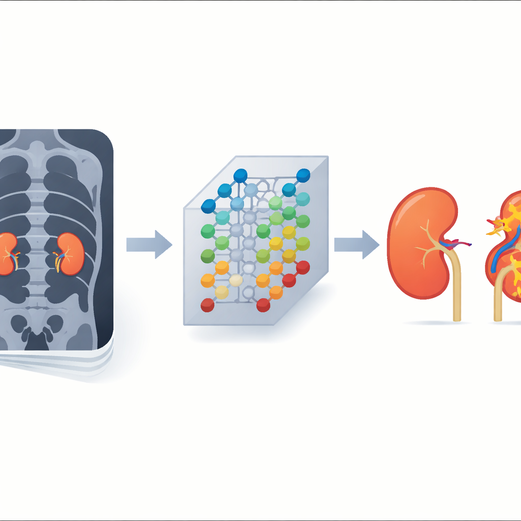
Teaching a computer to read subtle clues
To tackle this problem, the team collected CT scans and clinical data from 1661 patients treated at five hospitals plus a public dataset. They first built traditional “radiomics” models that measured many handcrafted features of the tumor and its surroundings, such as shape and texture. These models worked reasonably well but struggled to catch a large share of truly invasive tumors. The scientists then designed RENALNet, a three‑dimensional deep learning system that looks directly at the CT volumes of the tumor and the tissue ring around it, learning its own patterns rather than relying only on pre-defined measurements.
How the new tool performs with doctors
RENALNet was trained on part of the patient group and tested on the rest, as well as on four outside hospital cohorts to see how well it generalizes. Across these groups, the AI model was more sensitive than radiomics at detecting tumors that were secretly more advanced, while keeping accuracy high. Importantly, the researchers also asked junior, mid‑career, and senior radiologists to read the CT scans with and without the help of RENALNet’s risk scores. When the AI output was combined with each radiologist’s judgment, their ability to distinguish truly invasive tumors improved, especially for less experienced readers, showing how human expertise and AI can work together.
Connecting images to tumor behavior
The study went a step further by asking whether the AI’s risk predictions reflected real biological aggressiveness. In several patient groups, tumors that RENALNet labeled as high risk showed higher levels of Ki‑67, a marker of how quickly cancer cells are dividing. Among 246 patients with follow‑up data, those in the AI-defined high‑risk group were much more likely to see their disease progress within five years than those in the low‑risk group. Using gene activity data from a large public cancer program, the team found that high RENALNet scores lined up with activation of molecular pathways involved in invasion, inflammation, and survival of cancer cells, suggesting that the CT patterns the model uses are tied to deeper genetic programs inside the tumor.
What this could mean for care
Taken together, the findings suggest that RENALNet can act as a noninvasive window into how dangerous a kidney tumor really is, even when the CT scan looks deceptively calm to the naked eye. By flagging patients whose tumors are more likely to have already slipped beyond the kidney, the tool could help surgeons decide when it is safer to remove the whole kidney rather than attempt a smaller operation, and when closer follow‑up is warranted. While the model still needs to be tested in real‑time clinical practice and expanded to other scan types and tumor subtypes, it offers a promising example of how AI that “understands” both images and biology might sharpen cancer treatment decisions in the future.
Citation: Li, S., Wang, C., Li, F. et al. Noninvasive prediction of occult pT3a upstaging in localized ccRCC with radiogenomic insights and prognostic relevance. npj Precis. Onc. 10, 104 (2026). https://doi.org/10.1038/s41698-026-01315-2
Keywords: kidney cancer, medical imaging AI, deep learning, surgical planning, radiogenomics