Clear Sky Science · en
Integrated predictive model for visceral pleural invasion in small NSCLC with high clinical utility
Why this matters for people with lung cancer
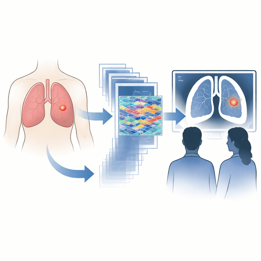
Lung cancer is still the deadliest cancer worldwide, and even very small tumors can behave very differently. One hidden warning sign is whether a tumor has broken through the lung’s smooth outer lining, a change that often calls for more aggressive surgery. This article describes a new computer-based tool that reads CT scans to flag this dangerous invasion more reliably, potentially helping doctors plan the right operation the first time.
Seeing beyond what the eye can catch
Doctors already use CT scans to look for signs that a lung tumor has reached the outer lining, such as subtle pulls on nearby tissue or delicate strands stretching toward the chest wall. But especially for small tumors, these clues can be faint and are interpreted differently by different radiologists. Yet this detail matters a lot: once the tumor crosses that boundary, its stage is raised and the risk of spread and recurrence jumps, even when the tumor measures less than three centimeters across. Patients with this kind of invasion usually need more extensive removal of nearby lymph nodes and closer follow-up, so missing it can change outcomes.
Blending three ways of reading a scan
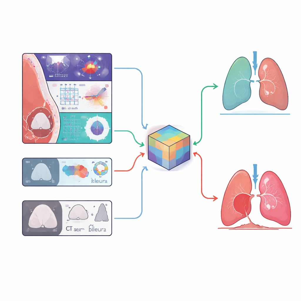
The researchers built what they call a multi-feature integrated imaging fusion, or MIIF, model to sharpen this decision. Instead of relying on one technique, they combined three kinds of information from pre-surgery CT scans of 2,822 small lung cancers collected at several hospitals. The first source was deep learning, in which a neural network learned complex patterns directly from three-dimensional image patches focused on the nodule and the lung surface. The second source, known as radiomics, captured hundreds of numeric descriptions of each tumor’s shape and texture that the human eye cannot easily quantify. The third source was a set of straightforward CT findings such as whether the nodule was solid or partly hazy, its solid core size, and how it touched or tugged on the pleura, the lung’s outer lining.
How well the tool performed
From these many measurements, the team used statistical methods to select 42 of the most informative features and trained a machine-learning classifier to estimate the chance of invasion for each tumor. When tested on patients who were not part of the training step, the combined MIIF model clearly outperformed a deep-learning-only model. In one hospital’s test group it showed excellent accuracy, and in an independent hospital it still reached an acceptable performance level, despite differences in scanners and imaging settings. The model was especially strong at correctly ruling out invasion, a key need when deciding whether a limited surgery is safe.
Helping radiologists make more consistent calls
The study also asked six chest radiologists, both senior and junior, to judge the same scans first on their own and later with the model’s risk estimate available. On average, the doctors’ accuracy and their ability to avoid false alarms improved when they could consult the MIIF output, with particularly large gains for less experienced readers. Their sensitivity, or ability to catch true invasion, stayed similar or improved slightly. This suggests that rather than replacing experts, the system works like a second pair of eyes that nudges borderline cases in a more consistent direction and narrows the gap between junior and senior judgments.
What the scan itself can still tell us
Alongside the computer model, the authors re-examined classic CT features linked to invasion. They found that purely hazy nodules showed no invasion in their data, while solid nodules were affected far more often than part-solid ones. Among tumors near the lung surface, a larger solid core, stronger pulling of the pleura, and certain attachment patterns were all independent warning signs. These are details that radiologists can continue to use in everyday practice, and they were among the human-understandable ingredients that fed into the MIIF model.
What this means for patients
In plain terms, this work shows that a carefully designed computer assistant can match seasoned specialists in spotting when a small lung tumor has already broken through the lung’s protective lining, and can lift the performance of the whole imaging team. If validated more widely and integrated into routine workflows, such a tool could help surgeons choose the right extent of surgery and lymph node removal, sparing some patients from undertreatment and others from unnecessarily aggressive operations. For people facing early-stage lung cancer, that could translate into more tailored care and better chances that the first operation is the one they really need.
Citation: Yang, S., Wei, Y., Wang, Q. et al. Integrated predictive model for visceral pleural invasion in small NSCLC with high clinical utility. npj Precis. Onc. 10, 97 (2026). https://doi.org/10.1038/s41698-026-01305-4
Keywords: lung cancer imaging, visceral pleural invasion, artificial intelligence in radiology, CT-based prediction, surgical planning