Clear Sky Science · en
Immunohistochemical biomarker-associated radiomics for classifying thymic epithelial tumors: a multicenter retrospective study
Seeing Hidden Clues in Routine Scans
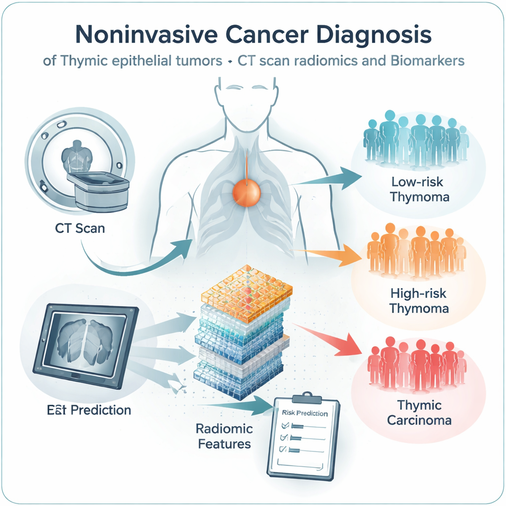
Doctors routinely use chest CT scans to spot tumors in the front of the chest, where a small organ called the thymus sits. But even experienced radiologists often struggle to tell which thymus‑related tumors are harmless and which are dangerous just by looking at the images. This study explores how advanced computer analysis of CT scans, combined with lab markers from tumor tissue, could offer a safer, noninvasive way to sort low‑risk from high‑risk tumors and guide treatment.
Why Tumors Near the Thymus Are Hard to Judge
Thymic epithelial tumors are the most common growths in the front part of the chest in adults. Some are relatively mild thymomas that grow slowly, while others are aggressive thymic carcinomas that invade nearby structures and spread. Today, doctors rely on standard CT images and a staging system to decide how serious a tumor is, but the pictures of different tumor types often look surprisingly similar. Biopsies can help, yet they carry extra risks in this tightly packed area near the heart and major blood vessels. Clinicians need better tools that can flag high‑risk cases early without adding danger or discomfort for patients.
Turning Pictures into Numbers
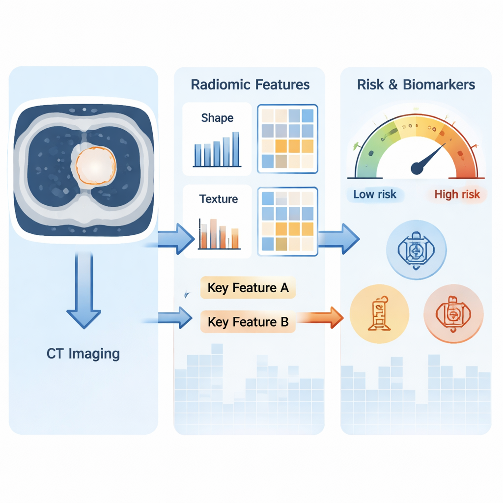
The research team worked with CT scans from 307 people with thymic tumors and 100 healthy volunteers from two hospitals. Using a technique called radiomics, they converted each tumor image into hundreds of numerical measurements that describe its shape, brightness, and texture in great detail—far beyond what the human eye can easily see. They then used computer algorithms to group tumors based on these patterns. Three distinct imaging groups emerged. One group was dominated by low‑risk thymomas, another mixed higher‑risk thymomas in both early and advanced stages, and a third consisted mainly of late‑stage thymic carcinomas. These imaging groups also lined up with important clinical features, such as stage of disease and lab test results from tumor samples.
Linking Image Patterns to Lab Markers
Pathologists often test thymic tumors for proteins like CD117 and TDT, which help distinguish more aggressive carcinomas from lymphocyte‑rich thymomas. The scientists asked whether the radiomic fingerprints in CT scans could predict these markers without needing tissue in hand. They found two standout image‑based measurements that tracked closely with CD117 and TDT levels. One captured how evenly the signal is spread across the tumor, and the other summarized the typical gray level in the image after a mathematical transformation. When they combined these two features into a simple score, they could reliably tell apart tumors that were CD117‑positive and TDT‑negative—typical of thymic carcinoma—from those with the opposite pattern, typical of thymoma. In multiple test groups, this score showed strong accuracy, suggesting it reflects real biological differences in how these tumors grow and organize their cells.
From Scores to Risk Predictions
The team then tested whether this image‑based score could do more than mirror lab tests—could it also predict how risky a tumor is overall? They compared the score against established measures of tumor aggressiveness, including whether a tumor fell into a low‑ or high‑risk category and whether it was in an early or advanced stage. In separate patient groups used for training and validation, the score performed well at signaling higher‑risk disease and more advanced stages, though it was less helpful for predicting unrelated factors such as age, sex, or the presence of myasthenia gravis, a nerve‑muscle disorder sometimes linked to thymic tumors. This pattern suggests that the radiomic features are tuned to the tumor’s core biology rather than general patient characteristics.
What This Could Mean for Future Patients
For someone facing a newly discovered mass near the thymus, the study’s message is hopeful: the CT scan you already need might someday provide much more than a simple picture. By automatically reading fine‑grained patterns in the images and tying them to known lab markers and risk groups, radiomics‑based tools could help doctors estimate how aggressive a tumor is and plan surgery or other treatments with greater confidence—potentially reducing the need for risky biopsies. While the authors note that their model still needs to be tested and standardized across more hospitals and scanners, their work points toward a future in which advanced image analysis becomes a routine, noninvasive companion to pathology in the care of thymic tumor patients.
Citation: Zhang, Y., Guo, Y., Li, J. et al. Immunohistochemical biomarker-associated radiomics for classifying thymic epithelial tumors: a multicenter retrospective study. npj Precis. Onc. 10, 73 (2026). https://doi.org/10.1038/s41698-026-01286-4
Keywords: thymic epithelial tumors, radiomics, CT imaging, cancer risk prediction, noninvasive biomarkers