Clear Sky Science · en
Innate lymphoid cell heterogeneity and etiology-specific reprogramming in hepatocellular carcinoma
Why liver cancer’s hidden defenders matter
Hepatocellular carcinoma, the most common form of primary liver cancer, kills hundreds of thousands of people each year. Yet tumors do not grow in isolation: they are surrounded by immune cells that can either fight the cancer or unintentionally help it. This study focuses on a little‑known group of immune cells, called innate lymphoid cells, and asks how they behave differently in liver cancers caused by chronic hepatitis B virus compared with those arising from non‑viral causes such as fatty liver disease or alcohol. Understanding these differences could point toward truly personalized immunotherapies for liver cancer patients.
A closer look at quiet immune residents
Innate lymphoid cells (ILCs) are rare sentinels that live permanently in tissues, including the liver. They react quickly to damage or infection by releasing powerful chemical signals, but unlike classic T cells they do not recognize specific pathogens. Because they are scarce and share markers with other immune cells, ILCs are almost invisible in standard bulk genetic analyses. The researchers overcame this by combining single‑cell RNA sequencing, high‑dimensional protein profiling (CyTOF), and bulk RNA sequencing on tumor and nearby non‑tumor liver samples from 50 patients. This allowed them to pick out ILCs one by one and determine which subtypes were present and what each cell was doing.
Many flavors of the same immune cell
The team discovered that liver ILCs are far from uniform. They identified several subgroups, including a proliferating, stem‑like version of group 1 ILCs (ILC1p), a strongly cell‑killing version (ILC1c), classic allergy‑associated group 2 ILCs (ILC2), and a rarer group 3‑like subset (ILC3). These groups could be distinguished by patterns of genes linked to growth, inflammation, and cytotoxic weapons such as perforin and granzymes. In healthy‑adjacent liver tissue from both hepatitis B and non‑viral patients, the balance of these subtypes looked broadly similar, suggesting that the basic ILC toolkit is shared. But when the researchers examined tumor tissue, the mix of subtypes and their activity changed dramatically.
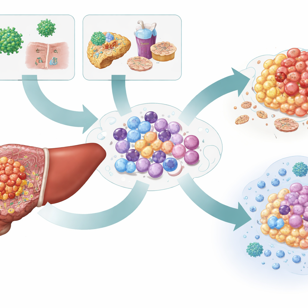
Viral and non‑viral tumors shape ILCs in opposite ways
In tumors driven by non‑viral causes, the proliferating ILC1p cells dominated and tended to mature into highly cytotoxic ILC1c cells and ILC2 cells. These ILC2s produced more IL‑13 and other factors tied to scarring and blood‑vessel growth, hinting that they may help build a fibrotic, tumor‑supporting niche. At the same time, ILC1c cells in non‑viral tumors expressed strong inflammatory and killing programs and responded to high levels of the cytokines IL‑12 and IL‑15 in the tumor microenvironment. Functional tests confirmed that these cells more often carried both interferon‑gamma and granzyme B, hallmarks of active tumor‑attacking cells.

Exhausted defenders in hepatitis B–related cancer
The picture was different in hepatitis B–associated tumors. There, ILC1c cells displayed more inhibitory receptors such as TIGIT and CD96, and gene signatures consistent with exhaustion rather than vigorous attack. Their communication with other immune cells also shifted. Instead of engaging CD8 T cells through activating contacts, ILC1c cells in viral tumors more frequently interacted through a brake‑like pathway involving the molecule HLA‑E and the CD94:NKG2A receptor, which is known to dampen T‑cell and natural killer cell activity. They also produced chemokines that can attract regulatory T cells and support tumor growth. Together, these signals point toward a more immunosuppressive environment in hepatitis B–related liver cancer.
What this means for future liver cancer care
To a non‑specialist, the key message is that not all liver cancers are equal in how they rewire the body’s rapid‑response immune cells. Non‑viral tumors seem to encourage a mix of ILCs that both fuel fibrosis and, under the right signals, can mount strong anti‑tumor attacks. Hepatitis B–driven tumors, in contrast, push ILCs toward a tired, inhibitory state that blunts immune defense. These findings suggest that patients might one day receive different immune‑boosting drugs based on how their cancer arose—for example, IL‑15–based therapies to amplify active ILC1c cells in non‑viral disease, or checkpoint blockers targeting inhibitory receptors in hepatitis B–related cancer. By mapping this hidden layer of immune diversity, the study moves the field closer to precision immunotherapy for liver cancer.
Citation: Lee, Y.H., Chuah, S., Leow, W.Q. et al. Innate lymphoid cell heterogeneity and etiology-specific reprogramming in hepatocellular carcinoma. npj Precis. Onc. 10, 122 (2026). https://doi.org/10.1038/s41698-026-01282-8
Keywords: liver cancer, innate lymphoid cells, hepatitis B, tumor microenvironment, immunotherapy