Clear Sky Science · en
A method for supervoxel-wise association studies of age and other non-imaging variables from coronary computed tomography angiograms
Why Heart Aging Patterns Matter
As people live longer, doctors are searching for better ways to tell how well a person’s heart is aging, beyond simple measures like blood pressure or cholesterol. This study looks inside detailed 3D X-ray scans of the heart to map how its shape and tissue properties change with age, and how those changes differ between women and men. By zooming in to very small subregions of the heart, the researchers hope to uncover early, hidden signs of aging that might eventually help predict disease risk or guide treatment.
Looking Inside the Beating Heart
The team worked with a special type of CT scan called coronary computed tomography angiography, which uses contrast dye to show the heart chambers and vessels in sharp detail. They analyzed scans from 1,388 volunteers aged 50 to 64 in the Swedish SCAPIS study, a large project designed to understand heart and lung health in the general population. For each person, the researchers had a 3D image of the chest along with basic information such as age and sex, but not detailed clinical diagnoses, allowing them to focus on broad patterns of aging in otherwise unselected adults.
From Raw Images to Comparable Hearts
Raw heart scans look different from person to person because of variations in body size, position, and anatomy. To compare subtle changes across hundreds of people, the researchers first used an automated tool to outline major heart structures such as the left and right pumping chambers, the upper chambers, the heart muscle, and the aorta. They then used a two-step registration process to “morph” every heart onto a common reference heart, ensuring that the same anatomical location lined up across all subjects. This deformable matching method was carefully checked using overlap scores and measures of how smoothly the images were warped, showing high accuracy for most people, especially on the left side of the heart.
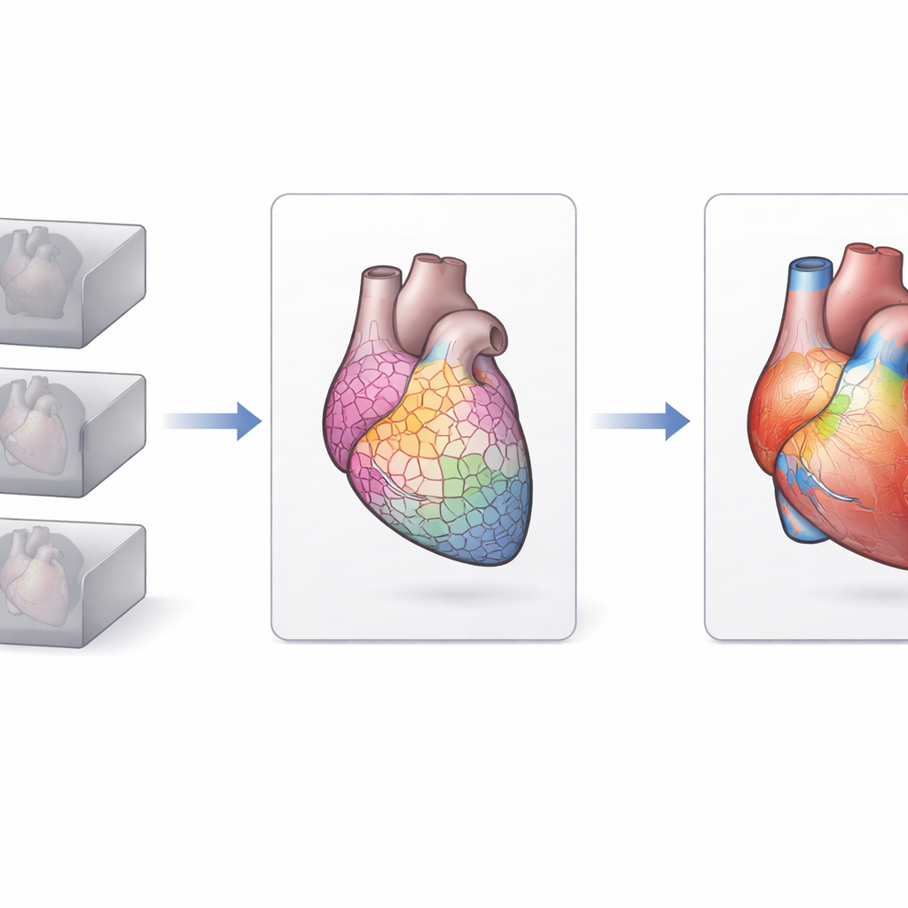
Dividing the Heart into Tiny Neighborhoods
Instead of studying only a few predefined regions, the authors divided the registered images into thousands of small, connected 3D patches called supervoxels. Each supervoxel behaves like a tiny neighborhood that averages the local volume changes and X-ray attenuation, a measure related to tissue density and contrast dye uptake. Working at this intermediate scale made the analysis faster and more robust, because it reduced random noise and the number of statistical tests. The team then used a technique called Imiomics, in which they calculated, for each supervoxel, how strongly its volume and attenuation were correlated with age across the entire cohort.

What Changes with Age in Women and Men
The supervoxel maps revealed both expected and new patterns of heart aging. In both sexes, parts of the left ventricle tended to be smaller in older individuals, in line with earlier studies showing that the main pumping chamber shrinks with age. In women, the left atrium—one of the upper chambers—tended to be larger with age, while this pattern was weaker and not clearly significant in men. The aorta showed local regions where volume increased with age in both sexes. The method also picked up changes outside the classic heart regions: fat around the coronary vessels showed a negative relationship with age, and certain bone marrow regions in the chest were linked to age in sex-specific ways. Many of these signals appeared in places that are not usually segmented or measured in routine analyses.
From Method to Future Health Insights
Overall, the study introduces a new way to turn rich 3D heart scans into detailed maps of how structure and tissue properties vary with age, separately in women and men. While the work does not yet provide direct diagnostic rules, it shows that supervoxel-wise analysis can uncover localized aging patterns that traditional region-based or black-box deep learning methods might miss or fail to explain. In the future, the same framework could be used to relate heart shape to biological age, risk factors, or specific heart diseases, helping clinicians move from a coarse, one-size-fits-all view of cardiac aging toward a more precise picture tailored to each part of the heart.
Citation: Öfverstedt, J., Lundström, E., Bergström, G. et al. A method for supervoxel-wise association studies of age and other non-imaging variables from coronary computed tomography angiograms. Sci Rep 16, 11000 (2026). https://doi.org/10.1038/s41598-026-46350-y
Keywords: heart aging, coronary CT angiography, cardiac imaging, supervoxel analysis, cardiovascular risk