Clear Sky Science · en
Precise disease heterogeneity and progression quantification in MSA and Parkinson’s disease using machine learning
Why this matters for people with movement disorders
People living with Parkinson’s disease or multiple system atrophy (MSA) often face years of uncertainty, because the two conditions can look very similar in the clinic while following very different paths. This study explores how advanced computer methods applied to brain scans might help doctors tell these diseases apart earlier, understand how they vary from person to person, and track how they change over time.
Looking inside the brain for clearer clues
Parkinson’s disease and MSA both involve abnormal buildup of a protein called alpha‑synuclein, but they damage different parts of the brain. Parkinson’s mainly affects a deep region that produces dopamine, while MSA attacks wider networks, including the cerebellum, brainstem, and movement circuits. On ordinary MRI scans these differences can be subtle, especially early on, and many people with MSA are initially misdiagnosed with Parkinson’s. The researchers used two kinds of MRI—structural scans that show brain shape and size, and diffusion scans that reveal the health of white matter wiring—to search for more precise, patient‑specific patterns.
Teaching computers to spot hidden patterns
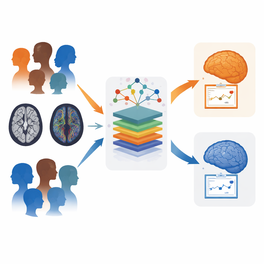
The team studied 17 healthy volunteers, 27 people with MSA (split into cerebellar and parkinsonian subtypes), and 15 with Parkinson’s, each followed for up to four yearly visits. They divided the brain into dozens of regions and measured local volume and two diffusion properties that reflect how water moves along nerve fibers. These measurements were fed into several machine‑learning models, which were trained to perform a simple task: decide whether a scan came from a person with MSA or with Parkinson’s. To avoid overfitting to such a rare disease, the authors used careful cross‑validation, repeated the training many times, and compared five different algorithm families before choosing the best performers.
From complex scans to a single personal score
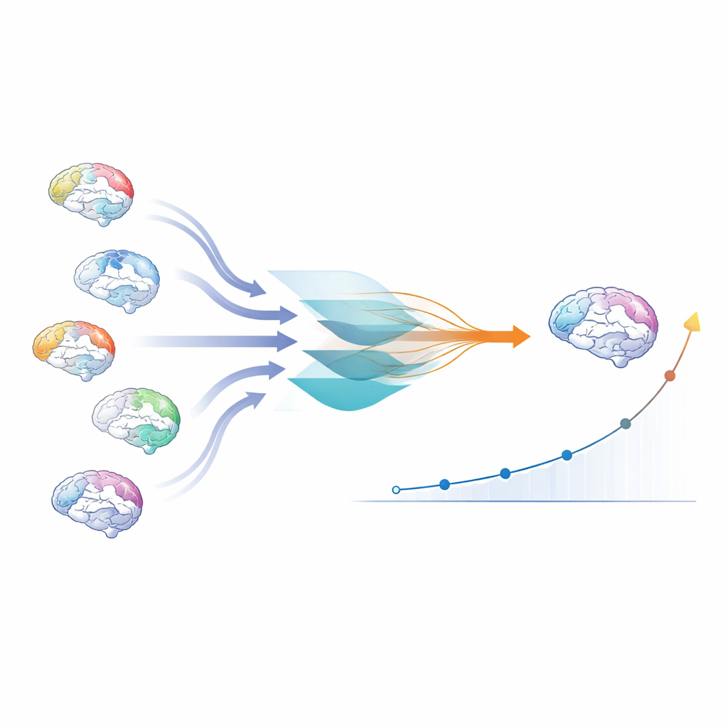
Rather than stopping at a yes/no computer diagnosis, the authors wanted a number that captured how strongly a person’s brain showed the signature of MSA versus Parkinson’s. They used an explainable AI method called SHAP, which breaks down each model decision into contributions from each brain region. These contributions served as weights, highlighting which areas the model found most informative. By combining the weights with the actual MRI measurements across all regions, they created three “heterogeneity” (HET) scores—one each for brain volume, and for the two diffusion measures. Each HET score boils a complex pattern of changes across the brain into a single summary value for each person and visit.
Seeing disease type and change over time
The new HET scores did more than just mimic existing MRI markers. They classified MSA versus Parkinson’s at least as well as, and often better than, a widely used atrophy index that focuses on a few key regions. Importantly, HET worked especially well for separating the parkinsonian form of MSA from Parkinson’s disease, a distinction that is notoriously difficult on standard scans. When the researchers looked across time, changes in the HET scores over one year tracked clinical worsening measured by a standard MSA rating scale better than simple measures of cerebellar shrinkage. Region‑by‑region maps of HET also recaptured known patterns of damage in MSA, such as degeneration of cerebellar and brainstem circuits, while revealing broader involvement of frontal and limbic white matter pathways and the connections between the brain’s two hemispheres.
What this could mean for patients and care
To a non‑specialist, the key message is that smarter analysis of routine MRI data can turn scattered signs of damage across the brain into a single, understandable score that reflects how “MSA‑like” a person’s pattern looks and how fast it is changing. This approach does not cure disease, and it still needs to be confirmed in larger groups, but it offers a promising tool for earlier and more accurate diagnosis, better tracking of progression, and more sensitive testing of new treatments in clinical trials. By honoring the fact that no two patients’ brains change in exactly the same way, the HET framework moves the field a step closer to truly personalized care in movement disorders.
Citation: Gebre, R.K., Raghavan, S., De Tora, M.E.J. et al. Precise disease heterogeneity and progression quantification in MSA and Parkinson’s disease using machine learning. Sci Rep 16, 10579 (2026). https://doi.org/10.1038/s41598-026-45949-5
Keywords: multiple system atrophy, Parkinson’s disease, brain MRI, machine learning, biomarkers