Clear Sky Science · en
Reduced cortical thickness in individuals with congenital adrenal hyperplasia (CAH)
Why hormone disorders can shape the brain
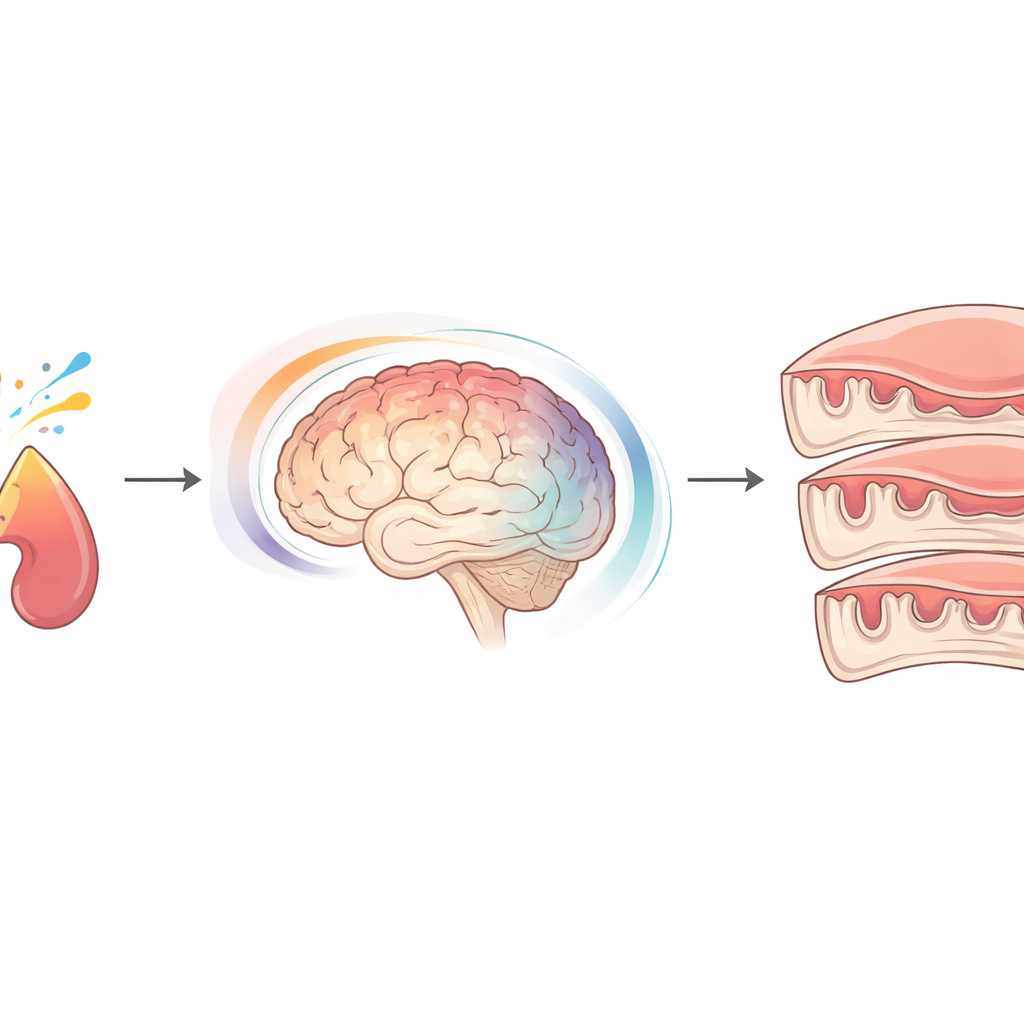
Many people have heard of hormones like cortisol, which helps the body handle stress, and androgens, often called “male” hormones. But fewer know that rare hormone disorders can subtly reshape the brain itself. This study looks at adults with congenital adrenal hyperplasia (CAH), a genetic condition that alters stress and sex hormones from before birth, to see whether their brain’s outer layer — the cortex — differs from that of people without the condition. Because the cortex is crucial for thinking, sensing, and emotions, even small changes in its structure could matter for brain health over a lifetime.
A lifelong hormone imbalance
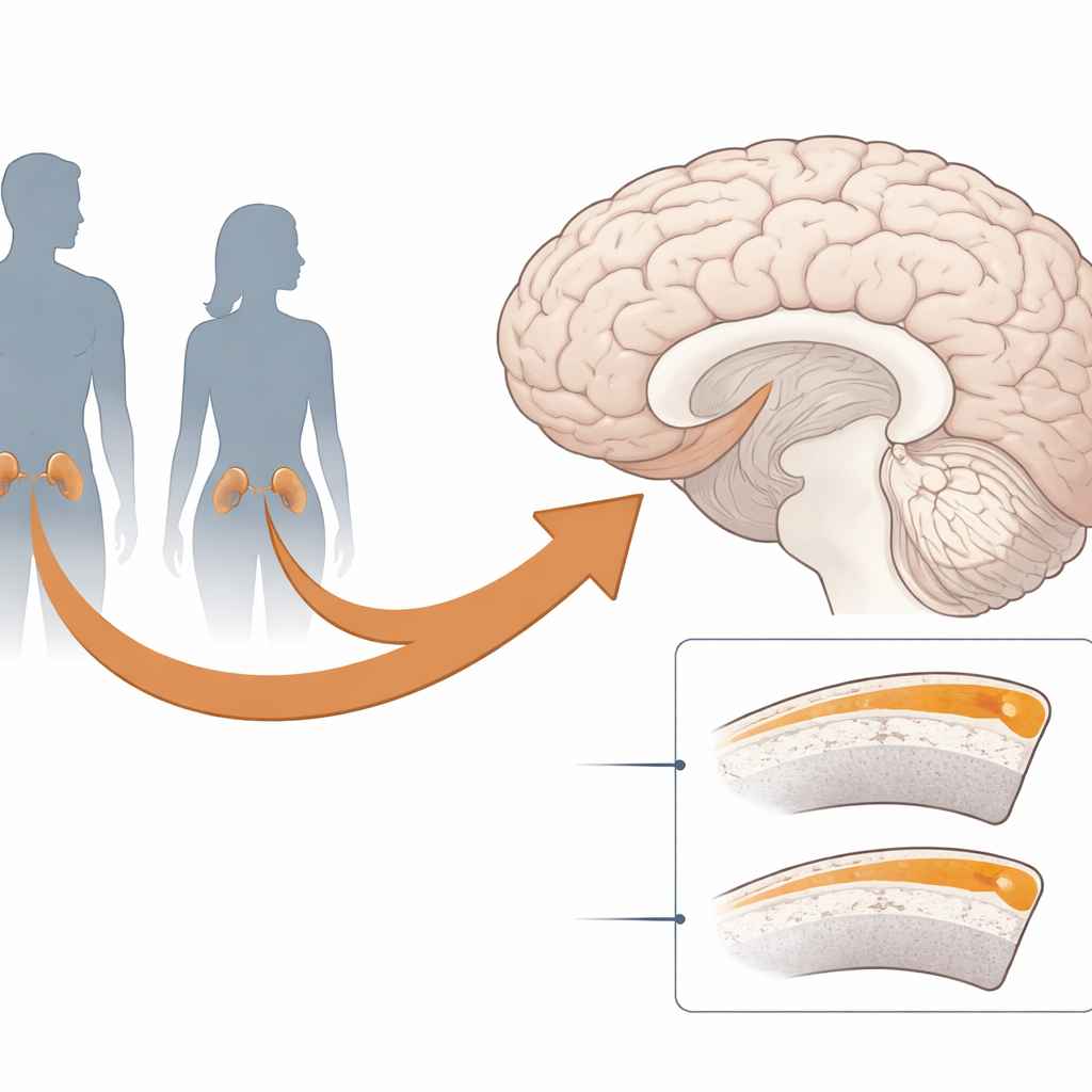
CAH is usually caused by a single faulty gene that prevents the adrenal glands from making enough cortisol. From early in pregnancy onward, affected fetuses have unusually low cortisol. In addition, girls with CAH are exposed to higher-than-typical levels of androgens, while boys have more typical androgen levels. After birth, people with CAH take daily hormone tablets to replace missing cortisol and, in girls, to bring androgens back into a typical range. Although this treatment is lifesaving, it may not perfectly mimic the body’s natural hormone rhythms, raising questions about how both the disorder and its treatment might influence the developing brain.
Scanning the thinking layer of the brain
The researchers focused on the cortex, the folded sheet of gray matter that sits on the brain’s surface and handles tasks such as movement, sensation, memory, and decision-making. They measured cortical thickness, which reflects the width of this sheet and is one common way to describe brain structure. Using high-resolution MRI scans and advanced surface-mapping techniques, they examined 53 adults with CAH (33 women and 20 men) and carefully matched them with 53 adults without CAH of the same age, sex, education level, and verbal ability. Instead of looking only at a few selected brain regions, they tested the entire cortex, point by point, while statistically controlling for the influence of age.
Where the cortex is thinner
The main finding was clear: on average, people with CAH had thinner cortices than their matched peers, and this pattern was widespread. Thinning appeared in both the left and right hemispheres, across the outer sides and inner surfaces of the brain, and in all four major lobes — frontal, parietal, temporal, and occipital. Some of the most affected areas help with planning and decision-making, touch and movement, and processing sights and sounds, such as regions in the frontal and parietal lobes, the precuneus, and parts of the temporal lobe. No brain region showed the opposite pattern; that is, there were no areas where the cortex was significantly thicker in CAH. Importantly, the differences were similar in women and men, and there was no sign that the effect of having CAH depended on biological sex.
How these results fit into the bigger picture
These findings build on earlier, smaller studies that also pointed to reduced gray matter in CAH, including changes in deep brain structures and specific parts of the cortex. With its larger sample and more sensitive methods, the current work revealed a broader pattern of cortical thinning than previously seen, and it echoed earlier results from the same group showing changes in white matter and in the large fiber bundle connecting the two hemispheres. Together, these studies suggest that CAH is linked not just to isolated spots of difference but to more global alterations in brain structure, detectable even in adulthood after years of hormone treatment.
What might be driving the brain changes?
The study cannot prove cause and effect, but it highlights several likely contributors. The genetic fault that causes CAH may influence the brain directly, beyond its impact on hormone levels. Low cortisol exposure before birth could alter how nerve cells grow, migrate, and connect, while long-term hormone replacement after birth, although essential, might sometimes expose the brain to higher or lower levels of glucocorticoids than ideal. On top of this, people with CAH often face ongoing physical and social stresses, from medical procedures to the daily demands of managing a chronic condition. The absence of sex-specific effects in the data suggests that extra prenatal androgens in girls with CAH are not the main driver of the observed cortical thinning.
What this means for people with CAH
For those living with CAH, these results do not mean that serious brain problems are inevitable, nor do they speak directly to day-to-day abilities. Instead, they show that the condition and its treatment leave a subtle, but widespread, fingerprint on the brain’s outer layer. Recognizing these structural differences may help doctors and researchers refine hormone therapies, monitor brain health more closely, and design future studies that link brain structure to thinking, mood, and quality of life. In simple terms, the work underscores that hormones shaping the body also quietly sculpt the brain, and that caring for people with CAH means caring for both.
Citation: Luders, E., Spencer, D., Hughes, I.A. et al. Reduced cortical thickness in individuals with congenital adrenal hyperplasia (CAH). Sci Rep 16, 9858 (2026). https://doi.org/10.1038/s41598-026-45407-2
Keywords: congenital adrenal hyperplasia, cortical thickness, brain MRI, hormones and brain, glucocorticoids