Clear Sky Science · en
Image quality and radiation dose of cone-beam CT versus multidetector CT for upper extremity osteosynthesis
Why clearer bone scans matter
When someone breaks a wrist or forearm badly enough to need a metal plate and screws, doctors rely on advanced X‑ray scanners to check how well the bone is healing and whether the hardware sits correctly. Two such scanners, cone‑beam CT and multidetector CT, are increasingly used for this purpose. This study asks a practical question of real concern to patients and clinicians alike: which type of scanner gives the clearer view of bone and metal implants, and how much radiation does each one deliver in the process?
Two ways to look inside a healing arm
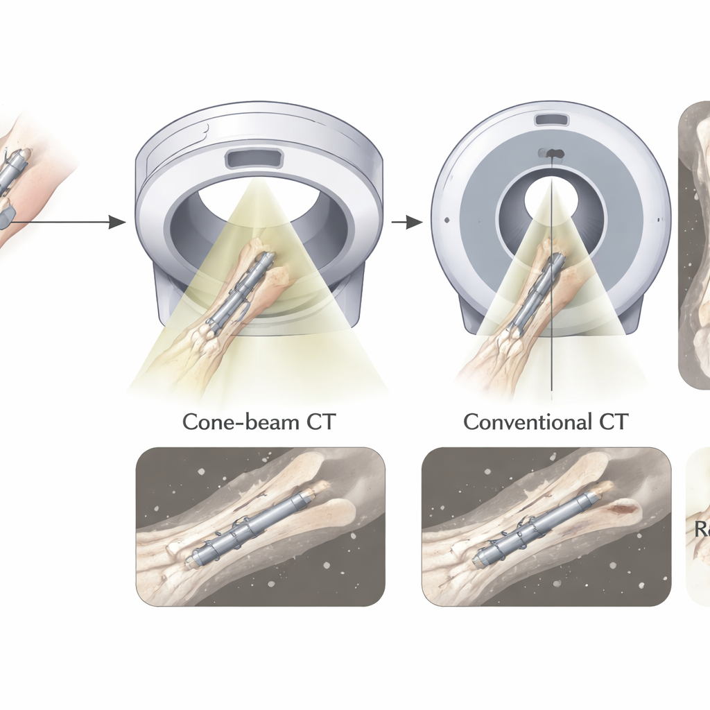
Both scanners create three‑dimensional pictures from X‑rays, but they do so in different ways. Cone‑beam CT uses a cone‑shaped beam and a flat detector that sweep around the limb in a single slow rotation, which makes the machines relatively compact and well suited for imaging arms and legs. Multidetector CT, the workhorse of hospital emergency departments, uses a narrow fan‑shaped beam that spirals through the body very quickly, capturing detailed slices that are later combined into a full volume. Each method has advantages and trade‑offs in terms of sharpness, sensitivity to metal hardware, and radiation dose.
A realistic test using a donor forearm
To compare the two techniques fairly, the researchers used a freshly preserved human forearm into which a surgeon had implanted a metal plate on the radius bone, mimicking a common wrist fracture repair. They placed tiny radiation sensors on the skin, near the bone and plate, and just outside the scan area to measure exposure. Then they performed 24 scans in total—twelve with each scanner—while carefully matching the main technical settings so that the comparisons would be as even as possible. Five radiologists, who did not know which scanner produced which images, rated how clearly they could see the hard outer shell of the bone, the spongy interior, and any disturbing streaks or distortions from the metal plate.
How dose and detail stacked up
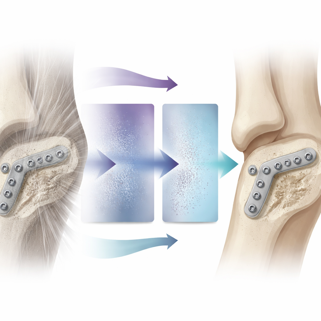
The radiation measurements showed that cone‑beam CT delivered a slightly higher overall dose along the scanned length of the forearm than multidetector CT. On average, the effective dose for cone‑beam CT was about a quarter higher. Yet both doses were extremely low compared with many everyday medical X‑rays and far below a year’s worth of natural background radiation, making the numerical difference unlikely to matter for an individual patient. Where the scanners truly parted ways was image quality. Both methods were equally good at showing the dense outer bone, but cone‑beam CT produced crisper views of the spongy bone inside and generated fewer bright streaks and shadows around the metal hardware. These impressions matched hard numbers: cone‑beam CT images had less random grain and more contrast between bone and surrounding tissue.
What this means for follow‑up care
The findings suggest that when the main goal is to inspect bone and metal plates in the wrist or forearm, cone‑beam CT can offer clearer pictures, especially of the delicate inner bone and the region right next to screws and plates, though at the cost of a modest increase in radiation. Multidetector CT, however, remains superior for situations where doctors must also examine soft tissues such as muscles and ligaments, or when scanning larger or heavier patients, thanks to its greater power and wider coverage. Because this study used a single donor forearm and focused only on bone, real‑world patients may show more variation, but the head‑to‑head design provides rare, carefully controlled evidence.
Balancing clarity and safety in bone imaging
For patients with repaired wrist fractures, this work indicates that both scanner types are safe and capable, but they are not interchangeable. Cone‑beam CT provides sharper bone views and cleaner images around metal implants, which can help doctors judge healing and hardware position with confidence. Multidetector CT, in contrast, offers slightly lower radiation and remains the first choice when a broader look at soft tissue injury is required. In everyday terms, the study shows that doctors can tailor the choice of scanner to the clinical question—prioritizing either the finest bone detail or the broadest overall picture—while keeping radiation exposure at very low levels in both cases.
Citation: Gökduman, A., Mahmoudi, S., Booz, C. et al. Image quality and radiation dose of cone-beam CT versus multidetector CT for upper extremity osteosynthesis. Sci Rep 16, 9719 (2026). https://doi.org/10.1038/s41598-026-44687-y
Keywords: cone-beam CT, multidetector CT, wrist fracture, radiation dose, bone imaging