Clear Sky Science · en
Multi-plane vision transformer for hemorrhage classification using axial and sagittal MRI data
Why this research matters for patients and doctors
When someone may be having a stroke or brain bleed, every minute counts. Brain scans can reveal dangerous hemorrhages, but reading these scans quickly and accurately is challenging, especially for magnetic resonance imaging (MRI), which produces many types of pictures in different viewing angles. This study introduces a new artificial intelligence (AI) method designed to read multi-angle MRI scans more like a skilled radiologist would, with the goal of spotting brain hemorrhages more reliably in real-world hospital conditions.
The challenge of finding brain bleeds on MRI
Intracranial hemorrhage—bleeding inside the skull—is a life-threatening emergency that demands rapid diagnosis. For decades, computed tomography (CT) has been the workhorse imaging tool for suspected brain bleeds because it is fast and relatively easy to interpret. MRI can match or even surpass CT in detecting hemorrhages and is better at showing how old the bleed is and revealing other problems such as areas of the brain that are starved of blood. However, MRI takes longer, is less widely available in some centers, and its images are more complex to interpret. This complexity makes it an attractive target for AI tools that can assist radiologists by screening large numbers of scans, flagging suspicious cases, and reducing the risk that a subtle but critical bleed is missed.
Why multiple views and scan types are hard for computers
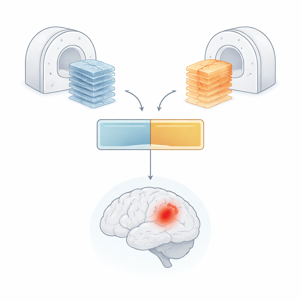
In routine clinical care, MRI of the brain is often acquired with relatively thick slices to keep the exam short, producing images that are much sharper in some directions than others. Radiologists view the brain in several planes—axial (top-down), sagittal (side-view), and sometimes coronal (front-view)—because some hemorrhages are easier to see from particular angles. Scans also come in several "contrasts" or flavors, such as FLAIR, diffusion, and susceptibility, each highlighting different tissue properties. Most current AI systems, however, expect all images to be lined up in a single standard orientation and at the same resolution. To meet this requirement, hospitals must digitally twist and resize the data, which can blur fine details and potentially hide small hemorrhages. Real clinical datasets add another complication: not every patient is scanned with the same set of contrasts, so models must cope with missing pieces of information.
A new multi-plane AI model that keeps more of the picture
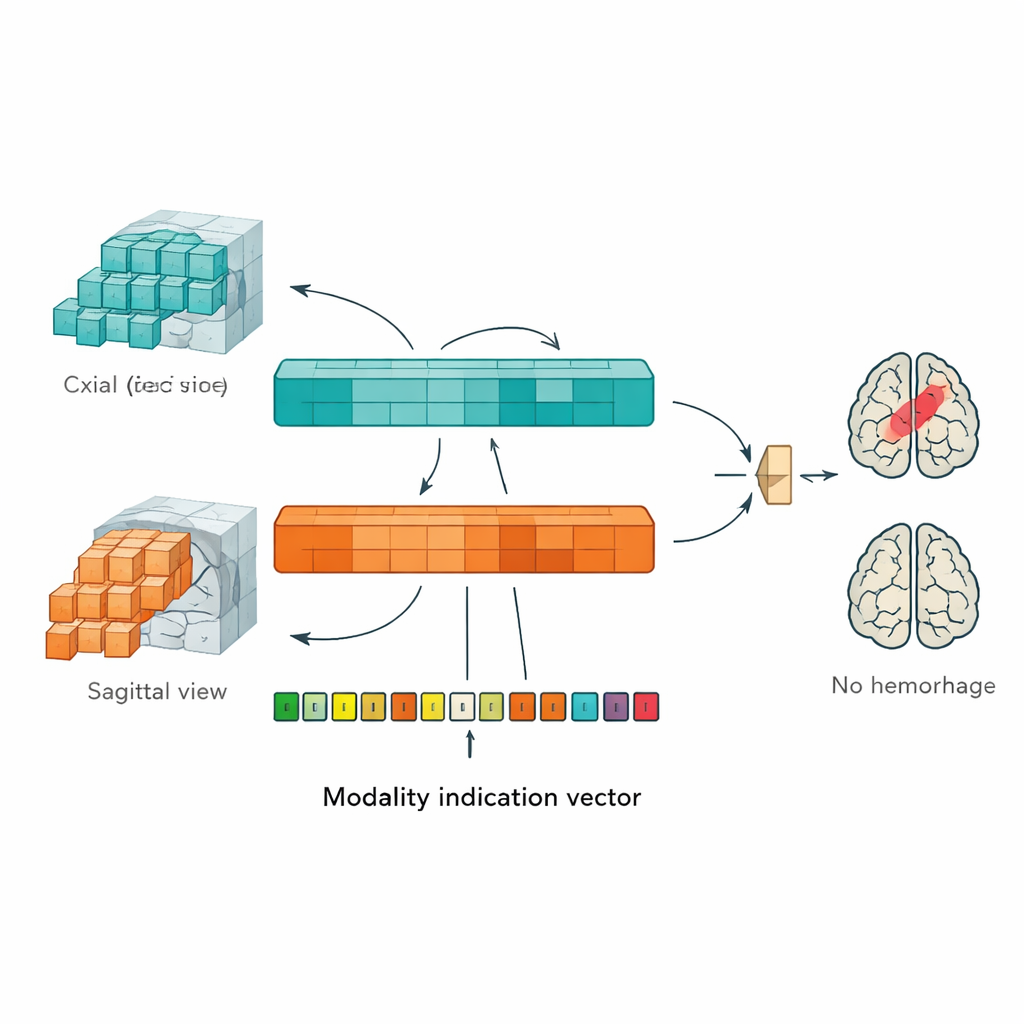
To address these problems, the authors designed a "multi-plane vision transformer" (MP-ViT), a type of AI originally developed for understanding natural images. Instead of forcing all MRI data into one viewing angle, MP-ViT has two dedicated processing branches: one for axial images and one for sagittal images. Each branch splits the three-dimensional brain into small blocks, converts them into tokens that the transformer can process, and then learns patterns that might indicate the presence of a hemorrhage. Crucially, these branches do not simply run in parallel and stay separate. The model uses a cross-attention mechanism to allow the two branches to exchange information, mimicking how a radiologist mentally combines views from different angles to form a clearer overall picture of the brain.
Handling missing scan types with a guidance signal
In real hospital workflows, not every patient has the same set of MRI contrasts; some may lack certain sequences such as special bleed-sensitive scans. To make the AI robust to these gaps, the authors added a "modality indication vector"—a simple code that tells the model which types of images are present and which are missing for a given patient. This vector is transformed into a set of internal signals that interact with the model’s learned features through an additional cross-attention step. In effect, the network is guided to adjust its expectations when certain kinds of information are unavailable, rather than being confused or overconfident. This design makes MP-ViT better suited to the messy, inconsistent data that arise in daily clinical practice.
How well the new method performs
The researchers trained and tested MP-ViT on a large, real-world dataset of over 12,000 MRI studies from three major scanner manufacturers, labeled by experienced radiologists as either showing acute or subacute intracranial hemorrhage or not. On an independent test set, MP-ViT achieved an area under the curve (AUC) of 0.854, a measure of how well it separates hemorrhage from non-hemorrhage cases across all possible decision thresholds. This score was notably higher than that of a standard vision transformer model working from a single plane, as well as several well-known convolutional neural network architectures such as ResNet and DenseNet. Statistical tests confirmed that these gains were unlikely to be due to chance. An internal analysis also showed that including the modality indication vector improved performance by more than one percentage point, underscoring the value of explicitly telling the model which scan types it has in hand.
What this could mean for future care
For a non-specialist, the key takeaway is that this study demonstrates a smarter way for AI to read MRI scans: it looks at the brain from more than one angle, keeps more of the original detail, and adapts when some types of images are missing. While the work was evaluated on a single internal dataset and focused only on classification rather than precise outlining of hemorrhages, it shows that carefully designed transformers can better match the messy reality of clinical imaging. If validated more broadly and integrated responsibly into hospital workflows, methods like MP-ViT could help radiologists detect brain bleeds more reliably in both emergency stroke settings and routine outpatient scans, potentially bringing faster treatment and safer outcomes to patients.
Citation: Das, B.K., Zhao, G., Mailhe, B. et al. Multi-plane vision transformer for hemorrhage classification using axial and sagittal MRI data. Sci Rep 16, 9333 (2026). https://doi.org/10.1038/s41598-026-44524-2
Keywords: brain hemorrhage, MRI, medical imaging AI, vision transformer, stroke diagnosis