Clear Sky Science · en
The quantitative and qualitative histomorphological structure of human stapes footplate
Why the smallest ear bone matters
Deep inside the skull, a structure thinner than a sheet of paper helps turn air vibrations into the sounds we hear. This study zooms in on the stapes footplate, the tiny plate where the last ear bone meets the inner ear. By mapping its fine structure in unprecedented detail, the authors show how this delicate part is built and why that matters when surgeons need to repair damaged hearing with tiny implants placed directly on it. 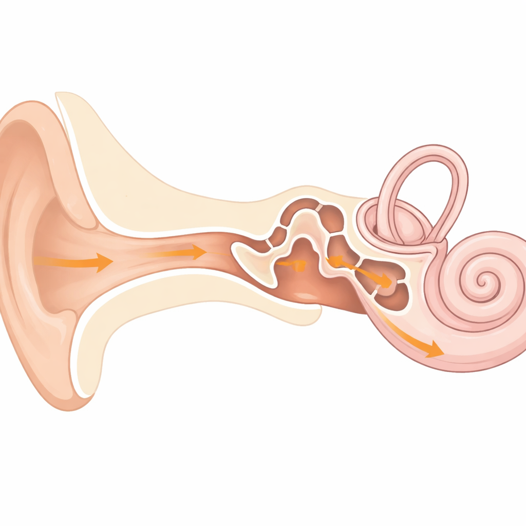
The last link in the hearing chain
Sound travels from the eardrum through three linked bones—the malleus, incus and stapes—before it reaches the fluid-filled inner ear. The stapes footplate is the contact surface between this mechanical chain and the inner ear. Even small changes in its shape or stiffness can weaken sound transmission. When disease destroys parts of the middle ear, surgeons often replace the bones with a metal prosthesis that presses on the footplate. To do this safely and effectively, they need to know exactly how thick the plate is and how its tissues are arranged.
Taking ultra-thin slices of a tiny plate
The researchers examined seven human stapes removed from donated temporal bones. After careful preparation, they cut the footplates into extremely thin slices—only one to two micrometers thick—and stained them to distinguish bone and cartilage. Using a microscope linked to analysis software, they measured tissue thickness and area at defined points across the plate, both along its length and across its width. In total, they collected about 1,400 measurements, allowing them to build a detailed map of how bone and cartilage are distributed in different regions.
Two layers with different jobs
The study revealed that the footplate is usually built as a two-layered structure: a bone layer facing the middle ear cavity and a cartilage layer facing the inner ear, covered by a thin lining of mucosa. In the central region, the total thickness averaged about one-tenth of a millimeter, with cartilage making up roughly three-fifths and bone two-fifths. Moving toward the edges where the footplate connects to its arch-like support, the plate became noticeably thicker—by more than a third overall. This thickening was driven mainly by an increase in bone, while the cartilage layer stayed relatively uniform. Seen from above, bone occupied a growing share of the plate’s area near the rim, suggesting that the outer regions are reinforced to bear higher mechanical loads.
Patterns, variability, and balance
Although the overall pattern—thin, more cartilaginous center and thicker, bone-rich edge—was consistent, the exact thickness of bone varied between individual footplates. In many spots, the total thickness stayed fairly constant while bone and cartilage traded places: where bone was thicker, cartilage tended to be thinner, and vice versa. In cross sections, the plate appeared more evenly thick from side to side, again with bone and cartilage working together. These findings point to a design in which bone sets the main shape and strength, while cartilage fine-tunes local stiffness and may help keep the plate’s surface and motion smooth and symmetric.
What this means for tiny ear implants
For surgeons, the most attractive place to press a prosthesis is the central region of the footplate, which couples well to inner ear motion. Yet this study shows that this very region has extremely thin bone—sometimes only a few micrometers thick—making it vulnerable to cracking if overloaded. At the same time, human ear bones appear to remodel very little in adulthood, limiting their ability to repair damage or grow firmly onto implants. These insights help explain why some traditional implants can cause fractures or leaks of inner ear fluid, and they support newer designs that spread out forces or use surface treatments to encourage more secure anchoring. 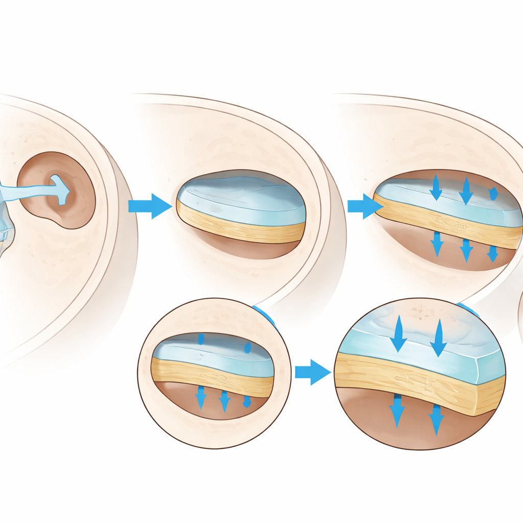
A clearer picture of a fragile gateway
By charting where bone and cartilage lie within the stapes footplate, this work provides a structural blueprint for improving hearing surgery. In simple terms, the plate’s center is a thin but important gateway for sound, while the edges are more heavily built to carry mechanical stress. Recognizing this balance can guide how and where tiny prostheses are placed, aiming to restore hearing without breaking the very structure they rely on.
Citation: Kemper, M., Türkeli, E., Kluge, A. et al. The quantitative and qualitative histomorphological structure of human stapes footplate. Sci Rep 16, 9537 (2026). https://doi.org/10.1038/s41598-026-43700-8
Keywords: middle ear, stapes footplate, hearing surgery, bone and cartilage, ossicular prosthesis