Clear Sky Science · en
Towards fully automated synthetic ECV quantification: an open-access machine learning-based approach for fast blood draw-free CMR
Why this matters for heart health
Doctors increasingly rely on heart MRI scans to spot early scarring in the heart muscle, a subtle change linked to many serious heart diseases. Today, getting this information typically requires both a time‑consuming image analysis and a blood test. This study shows that a computer can automatically read special heart MRIs and estimate the same information without drawing blood, opening the door to faster, more comfortable, and potentially wider access to advanced heart diagnostics.
Seeing hidden scars in the heart
Many chronic heart conditions cause fibrosis—tiny patches of scar‑like tissue that stiffen the heart muscle and worsen long‑term outcomes. Modern cardiac MRI can measure something called extracellular volume (ECV), which reflects how much of the heart muscle is taken up by fluid and fibrous tissue rather than healthy cells. ECV has become a powerful imaging marker for diffuse fibrosis, but measuring it in practice is cumbersome. It usually requires manual drawing of regions on several MRI images, careful correction of motion, and a recent blood test to determine hematocrit, the proportion of red blood cells in the blood.
The problem with blood tests and manual work
In real‑world hospitals, this traditional workflow is a bottleneck. Not all centers can collect a blood sample right around the time of the MRI, and hematocrit itself can fluctuate with factors as simple as body position. The image analysis also depends on trained experts, specialized software, and several manual steps where people outline heart structures slice by slice. These steps take time and can vary from one reader or center to another, which makes it harder to compare results across hospitals or large research studies.
Teaching a computer to read heart maps
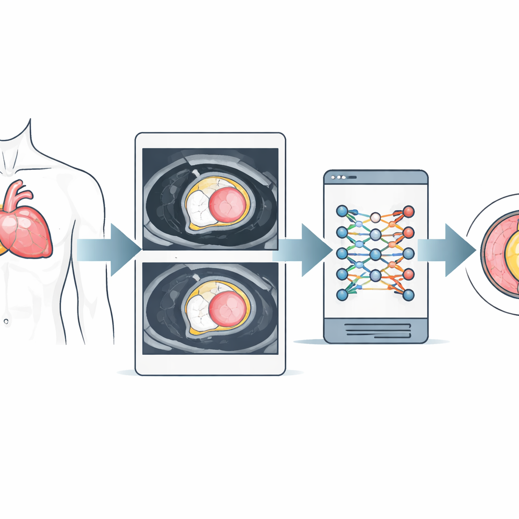
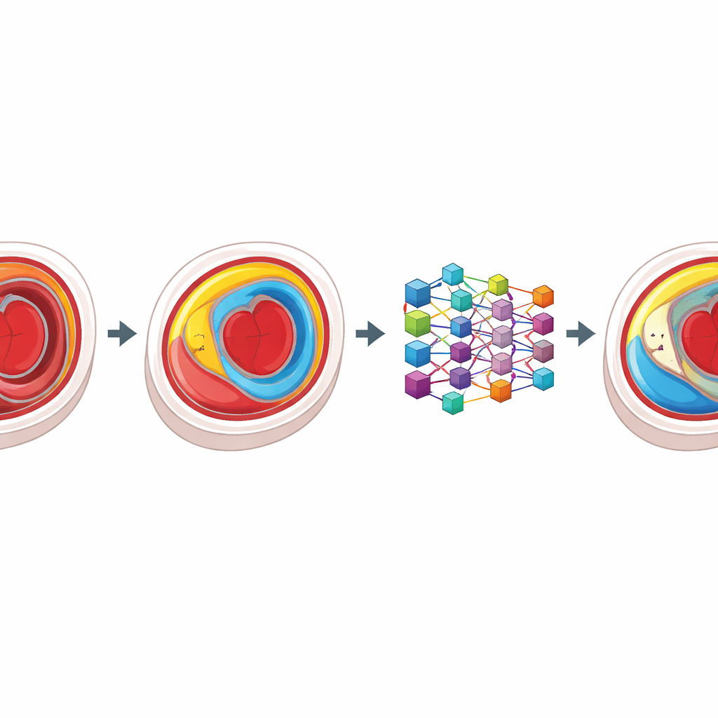
Building on earlier work that showed hematocrit can be estimated directly from MRI signal in the heart’s blood pool, the authors set out to automate the rest of the process. They used MRI data from over 1000 patients scanned at two different magnetic field strengths. In the training phase, experts carefully outlined the borders of the heart muscle and blood chambers on special T1 “maps” taken before and after contrast injection. These examples were used to train a deep‑learning model (a U‑net–type neural network) to find the same structures automatically. The algorithm also applied image‑processing steps to separate blood from nearby tissue and slightly shrink the borders to avoid blurring from motion.
From raw scans to synthetic ECV in one step
Once trained, the model was tested on a separate group of patients. For each person, it automatically measured typical T1 values in the heart muscle and blood, plugged those values into published formulas that estimate hematocrit from MRI alone, and then calculated a “synthetic” ECV—completely without a blood draw or manual contouring. The researchers compared this fully automated synthetic ECV with the conventional ECV that had been calculated using expert contours and lab‑measured hematocrit. Overall, the two methods agreed closely: the average values were almost identical, and the correlation between them was strong. Agreement was particularly good in the clinically important range up to about 35% ECV, where most patients fall.
What worked well and where it struggles
The automated contours were generally rated acceptable to excellent by independent heart imaging specialists, and the model handled images from both MRI field strengths reliably. However, differences between the automated and conventional measurements grew larger at very high ECV values, a range that often reflects severe disease. The authors suggest this is partly because such extreme cases were rare in the dataset, and partly because image quality and tricky anatomy can confuse the algorithm. They also note that the traditional reference uses a small region in the heart’s septum, while the automated method averages the entire slice, which naturally introduces some differences.
What this means going forward
For now, this approach is best viewed as a research tool rather than a ready‑made clinical replacement. Still, it shows that a computer can take standard pre‑ and post‑contrast heart MRI maps and, with no blood test and minimal human input, produce ECV values that closely track today’s labor‑intensive measurements in most patients. Because the code and trained model are openly available, other centers can test, refine, and adapt the method to their own scanners. If further validated—especially in patients with very high ECV—fully automated, blood‑free ECV could make advanced fibrosis assessment faster, more consistent, and more widely available.
Citation: Beyer, R.E., Hüllebrand, M., Doeblin, P. et al. Towards fully automated synthetic ECV quantification: an open-access machine learning-based approach for fast blood draw-free CMR. Sci Rep 16, 8552 (2026). https://doi.org/10.1038/s41598-026-43624-3
Keywords: cardiac MRI, myocardial fibrosis, extracellular volume, deep learning, medical imaging automation