Clear Sky Science · en
Increased brightness of fluorescent uridine, qU, inside single- and double-stranded RNA
Why Making RNA Glow Matters
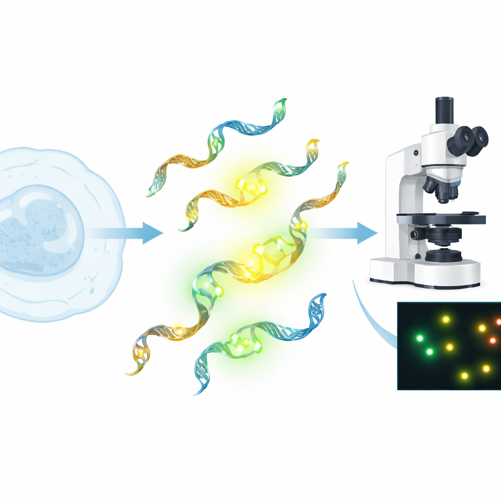
RNA sits at the heart of how our cells work and how many new medicines are being designed, from vaccines to cutting-edge genetic therapies. To truly understand what RNA is doing inside a cell—where it goes, how it folds, and how it interacts with other molecules—researchers need ways to make it light up without disturbing its natural behavior. This study presents a new glowing building block, a modified version of the natural RNA letter uridine called qU, that becomes exceptionally bright when built into RNA strands, opening the door to clearer, more precise imaging of RNA in action.
A New Way to Light Up RNA
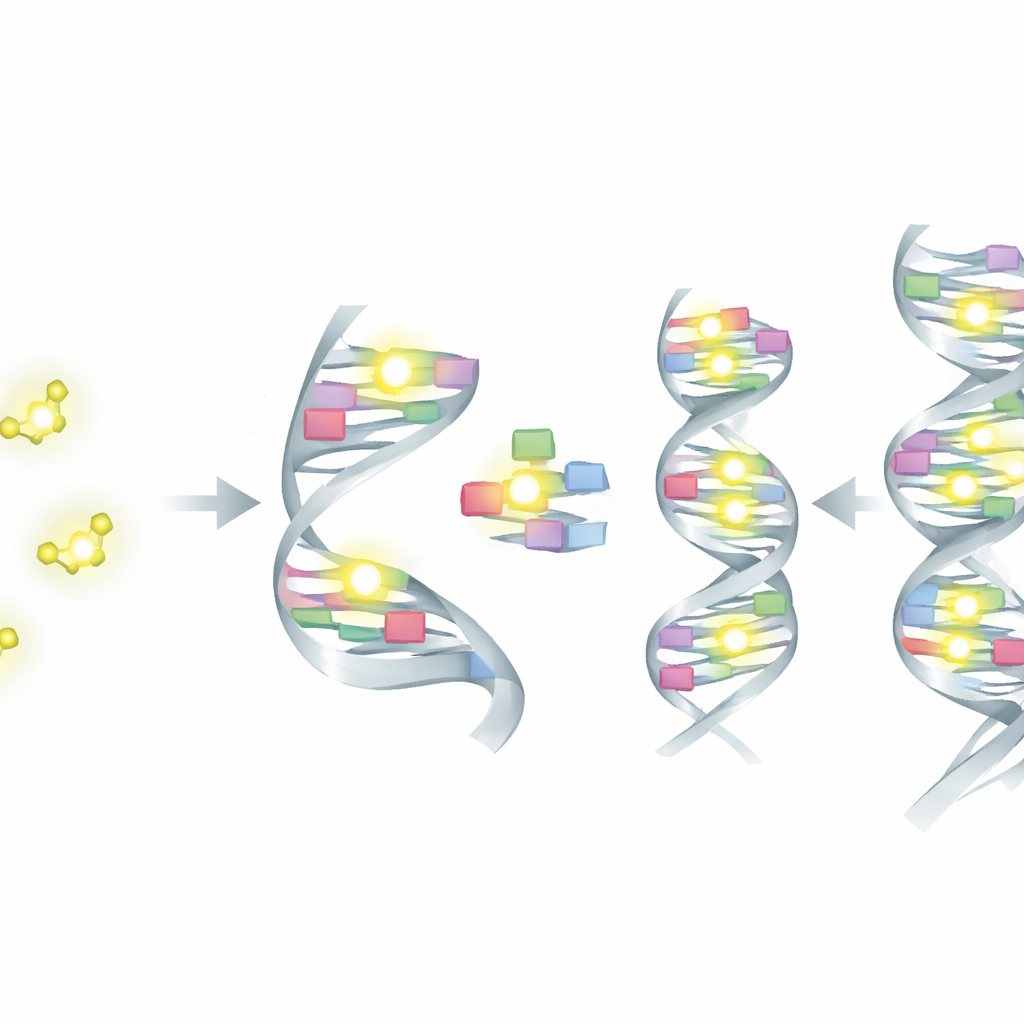
Traditional fluorescent dyes used to track RNA are usually attached on the outside of the molecule and are chemically very different from RNA itself. While bright, they can alter how RNA behaves, potentially changing how it folds, binds partners, or moves inside cells. In contrast, “fluorescent base analogues” mimic the natural letters of RNA and sit directly inside the genetic stack, offering a subtler way to label RNA. The authors focus on a new such analogue, quadracyclic uridine (qU), which had previously shown promising brightness when free in solution. Here, they ask: what happens to its glow and to RNA’s structure when qU is actually built into real RNA strands?
Building the Glowing RNA Pieces
To answer that, the team first developed a multi-step chemical route to convert qU into a special form (a phosphoramidite) that can be used in standard automated RNA synthesis. Using this, they created short RNA pieces where one normal uridine was replaced by qU and systematically varied the neighboring letters around it. They then combined these qU-bearing strands with matching partner strands to form double helices, or with slightly mismatched partners, and compared them to unmodified RNA. Along the way, they used a suite of optical techniques—including absorption and fluorescence measurements, lifetime analysis, RNA melting experiments, and circular dichroism—to see both how brightly qU glows and how much it disturbs the native RNA shape.
Brighter Glow Inside Real RNA
One of the most striking findings is that qU actually becomes brighter when placed inside RNA, in both single strands and double helices. Many fluorescent bases dim once they are surrounded by other bases; qU does the opposite. Its fluorescence efficiency rises from about one quarter when free in solution to as high as roughly two-thirds when part of an RNA strand, making it one of the brightest uridine-like labels reported so far. The exact brightness and how long it stays excited depend on the neighboring letters and whether the strand is single or double, showing that qU is sensitive to its local microenvironment. This sensitivity could be useful for reporting subtle structural changes or pairing mismatches along an RNA.
How the Glow Affects RNA Structure
Brightness, however, comes with a trade-off. When qU replaces a natural uridine in an RNA double helix, the helix becomes less stable: its melting temperature, a measure of how easily the two strands separate, typically drops by about 9 degrees Celsius. Spectroscopic fingerprints suggest that qU mostly adopts a form (called the iminol form) that does not pair perfectly with adenine, the base that uridine normally matches. This imperfect pairing likely increases local “breathing” or base flipping, mildly loosening the helix around the modified site. Despite this destabilization, circular dichroism measurements show that the overall helical shape of the RNA remains the usual A-form, meaning that the global architecture of the molecule is preserved even though local stability is reduced.
Using pH and Pairing to Tune the Signal
The authors also explored how acidity and base pairing partners influence qU’s glow. Just like the free molecule, RNA-bound qU responds strongly to changes in pH, especially under basic or acidic conditions, where its brightness and fluorescence lifetimes drop and its color shifts. This makes qU a potential reporter of local pH changes, such as those that occur when RNA enters acidic cellular compartments during uptake. Intriguingly, when qU faces mismatched partners instead of its usual adenine opposite, its brightness can become even higher than in correctly paired helices, and some of these mismatches actually stabilize the duplex compared to natural RNA with the same mismatch. This suggests that qU can probe both correct and incorrect pairing events while remaining highly emissive.
What This Means for Future RNA Studies
In everyday terms, this work delivers a powerful new “light bulb” that can be built directly into RNA’s text without rewriting its overall shape. Although replacing a single base with qU slightly weakens local pairing, the global helix remains intact, and the exceptional brightness—combined with strong response to its surroundings—makes qU an attractive internal label for demanding experiments, including fluorescence microscopy and lifetime imaging inside cells. Strategically placing qU in flexible or non-paired regions of RNA could allow researchers to follow therapeutic RNAs, watch structural rearrangements, and study binding events with high clarity, all while keeping the RNA as close as possible to its natural form.
Citation: Karlsson, A.F.E., Pfeiffer, P., Le, HN. et al. Increased brightness of fluorescent uridine, qU, inside single- and double-stranded RNA. Sci Rep 16, 8481 (2026). https://doi.org/10.1038/s41598-026-43188-2
Keywords: fluorescent RNA label, uridine analogue, nucleic acid imaging, RNA structure, fluorescent base analogue