Clear Sky Science · en
Prevalence and management practices of ophthalmic lesions in laboratory mice
Why tiny eyes matter in big science
Millions of laboratory mice are used worldwide to study diseases and test new treatments, and their eyes quietly play a double role: as research tools and as windows into animal wellbeing. This study asks a deceptively simple question: how often do people working with lab mice really look at their eyes, what do they find, and what do they do about it? The answers reveal that eye problems are common, often painful, and surprisingly easy to miss—yet better, routine checks could both improve animal welfare and make research results more reliable.
How the study took a closer look
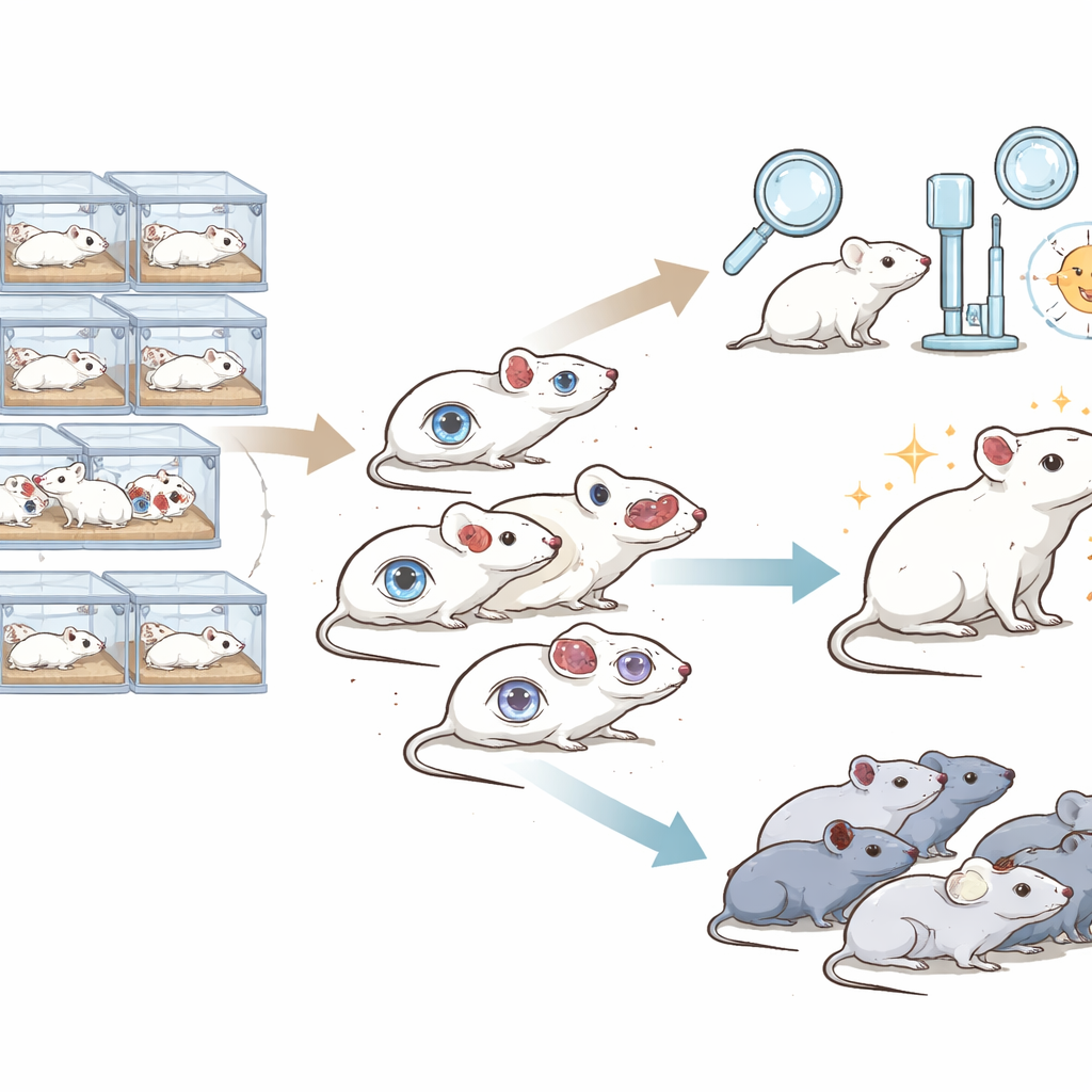
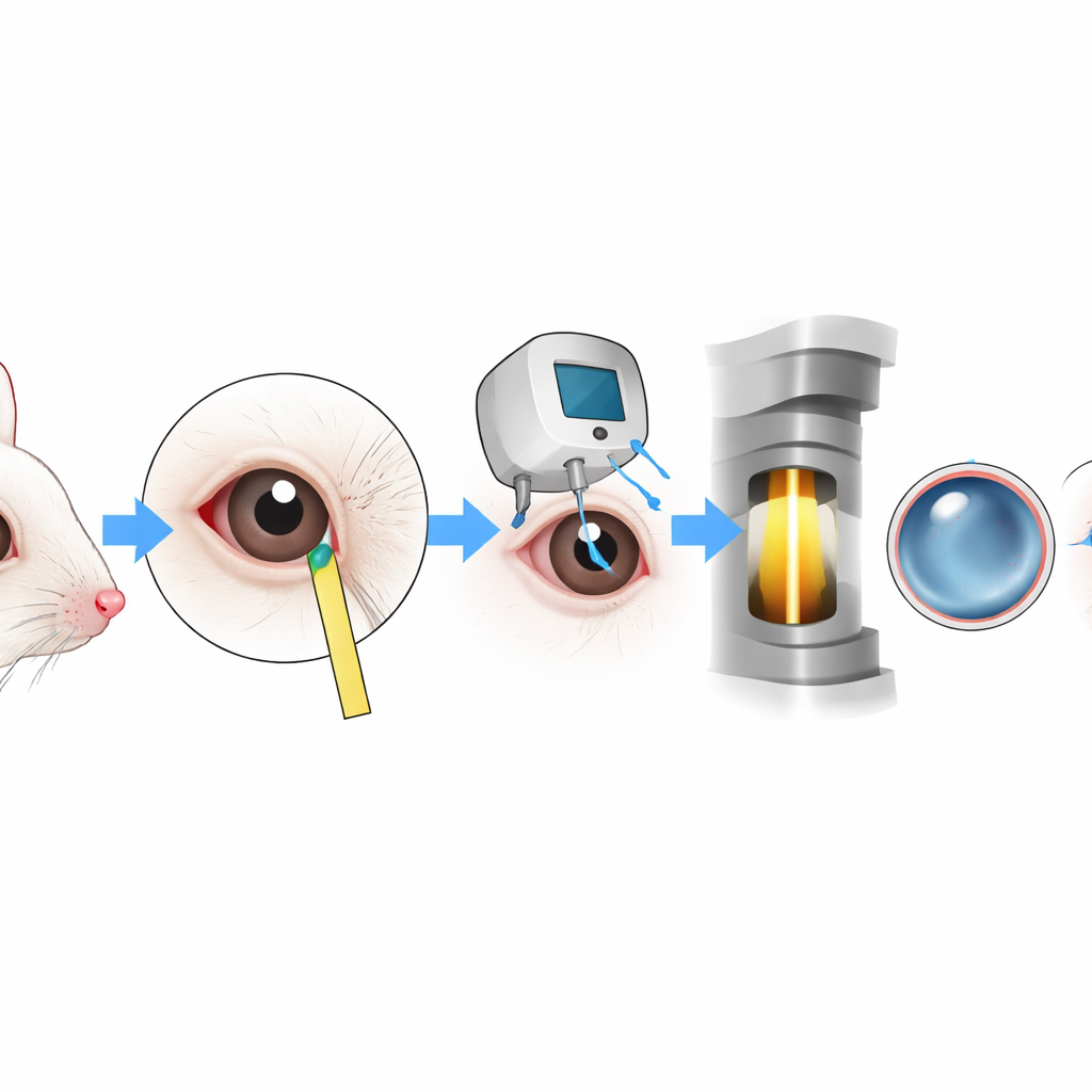
The researchers combined two approaches. First, they sent an online questionnaire to veterinarians, animal technicians, and other staff who care for mice in research facilities across Germany, Austria, and Switzerland. These 128 participants reported how eye changes are noticed, which problems they see most often, what diagnostic tools they use, and whether they try to find underlying causes. Second, in one large facility housing about 10,000 mice, the team systematically examined 142 individual animals flagged by caretakers for “any eye abnormality,” using magnification, special dyes, pressure measurements, and tissue analysis to understand exactly what was going wrong in the front part of the eye.
What people see—and what they miss
The survey revealed that most eye changes are picked up casually during routine cage changing, not through dedicated eye checks. Over 80% of respondents said that special eye examinations were done never or only in rare cases, and only about 14% reported that eye problems were regularly analyzed for their root causes. While many facilities could send samples to outside laboratories, only a small minority had slit lamps, pressure gauges, or simple dye tests readily available in-house. Staff frequently reported lens clouding, small or missing eyes, inflamed eyelids and conjunctiva, and injuries to the eyelids, but in roughly 70% of cases nobody systematically investigated why these lesions occurred.
What careful examination uncovered
When the researchers applied a structured examination scheme to the 142 affected mice, they found a broad spectrum of eye disease that routine inspection alone often could not distinguish. Most cases involved cloudy corneas, sometimes with ulcers, new blood vessel growth, or thickening of the tissue. Some mice had cataracts in the lens that were only reliably diagnosed with a slit lamp or under the microscope; without these tools, such changes could easily be confused with corneal haze. Others showed small or underdeveloped eyes (microphthalmia), narrowed eyelid openings, and sticky secretions. Swabs from eyes with obvious discharge grew common skin bacteria such as staphylococci and streptococci, suggesting that environmental factors or pre-existing surface damage can pave the way for infection even in high‑health laboratory colonies.
Why these findings matter for animals and experiments
The front of the eye, especially the clear cornea, is densely supplied with nerve endings and is highly sensitive to pain. Histological sections from affected mice showed thickened, inflamed tissue, new blood vessels, and, in severe cases, pus filling the front chamber of the eye—changes that are very likely to be painful or at least deeply uncomfortable. The study notes that some eye conditions in mice are often dismissed as harmless “background lesions,” tied to strain genetics or aging. Yet many of the observed problems, particularly surface injuries and infections, can cause pain, alter behavior, and potentially confound research that depends on normal vision, such as navigation or social interaction studies. The authors also show that simple measurements of tear production and eye pressure can detect functional disturbances and help differentiate between types of disease.
Building better routines for eye care
Overall, the work argues that healthy eyes should be treated as a basic requirement, not an optional extra, in laboratory animal care. The authors propose a straightforward examination pathway that starts with simple visual inspection and magnified images, then adds targeted tests—such as dye staining, pressure measurement, and slit-lamp evaluation—when problems are suspected. They recommend that facilities invest in basic ophthalmic tools, train staff to recognize and classify eye lesions, and avoid automatically blaming genetics without proper diagnosis. For animals with severe developmental eye defects or chronic painful conditions, humane removal from breeding and experiments, including euthanasia when necessary, is advised.
What this means going forward
To a lay reader, the message is clear: even in highly controlled research settings, mice can suffer from overlooked eye disease, and simple, systematic checks can make a big difference. By taking mouse eyes seriously—distinguishing between harmless quirks and painful injuries, and treating problems rather than ignoring them—scientists can both reduce suffering and strengthen the quality of their own data. In other words, better eye care for mice is not just an ethical upgrade; it is also a scientific one.
Citation: Matzek, D., Rumpel, S., Kassumeh, S. et al. Prevalence and management practices of ophthalmic lesions in laboratory mice. Sci Rep 16, 8732 (2026). https://doi.org/10.1038/s41598-026-43181-9
Keywords: laboratory mice, eye disease, animal welfare, corneal lesions, research ethics