Clear Sky Science · en
Clinical, radiological and laboratory features and outcomes of diffuse alveolar hemorrhage
When the Lungs Start to Bleed
Most of us think of lung problems as infections like pneumonia or chronic conditions like asthma. But there is a far rarer and more dramatic emergency in which the tiny air sacs of the lungs suddenly fill with blood, drowning out the very space where oxygen should enter the body. This condition, called diffuse alveolar hemorrhage, is difficult to diagnose, often tied to complex immune diseases, and can quickly become life-threatening. The study summarized here looks closely at who develops this syndrome, what it looks like on scans and lab tests, and which early warning signs are linked to survival in two major hospitals in Bogotá, Colombia. 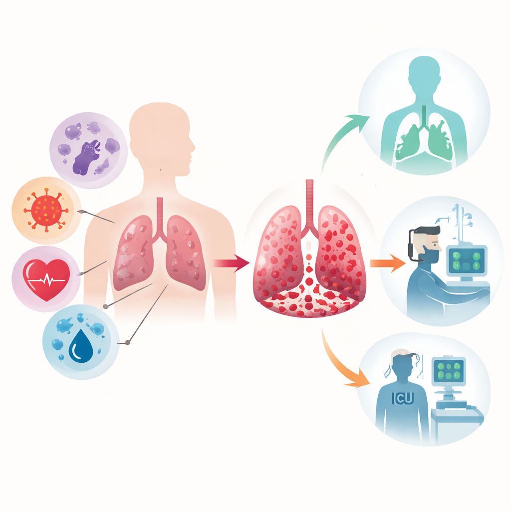
What This Dangerous Lung Bleeding Is
Diffuse alveolar hemorrhage occurs when fragile blood vessels surrounding the lung’s air sacs rupture, allowing blood to spill into spaces meant for air. People usually arrive at the hospital short of breath, coughing, sometimes bringing up blood, and often with low oxygen levels. Because these symptoms can mimic severe pneumonia or other lung diseases, doctors need special tools to confirm that bleeding is the core problem. One key test is bronchoalveolar lavage, in which a doctor rinses part of the lung with saltwater during a bronchoscopy and then examines the recovered fluid for blood-filled immune cells. In this study, only adults whose lavage fluid clearly showed these cells were included, ensuring that all 95 cases truly had this form of lung bleeding.
Who Was Affected and Why
The researchers found that patients were, on average, about 50 years old, and men and women were roughly equally affected. The striking finding was that four out of five cases were driven by autoimmune diseases—conditions in which the body’s own defense system attacks its tissues. Many of these patients had a form of blood-vessel inflammation known as ANCA-associated vasculitis, while others had illnesses such as lupus or related immune disorders. A smaller share of cases came from infections, heart failure, blood cancers, or complications of blood-thinning drugs, and a few had no clear cause. Most patients were already immunocompromised, either because of the disease itself or because they were taking strong immune-suppressing medications, which may make them both vulnerable to the bleeding and to secondary infections.
What Doctors Saw on Scans and in the Lab
On chest CT scans, nearly all patients had hazy, cloud-like areas called ground-glass opacities that covered both lungs—an imaging pattern that, while not unique, strongly raises suspicion for this type of bleeding when paired with the right symptoms. Lab tests showed that anemia was common and that many patients had antibodies linked to autoimmune disease or changes in complement proteins, which are part of the immune system. The bronchoalveolar lavage samples were rich in blood-laden macrophages, confirming ongoing bleeding. Most patients received high-dose steroids to calm the immune system, and many also got other strong treatments such as cyclophosphamide, plasma exchange, or rituximab. Supportive care included dialysis for kidney failure, antibiotics when infection was suspected, and adjustments to blood thinners in those whose medications contributed to the problem. 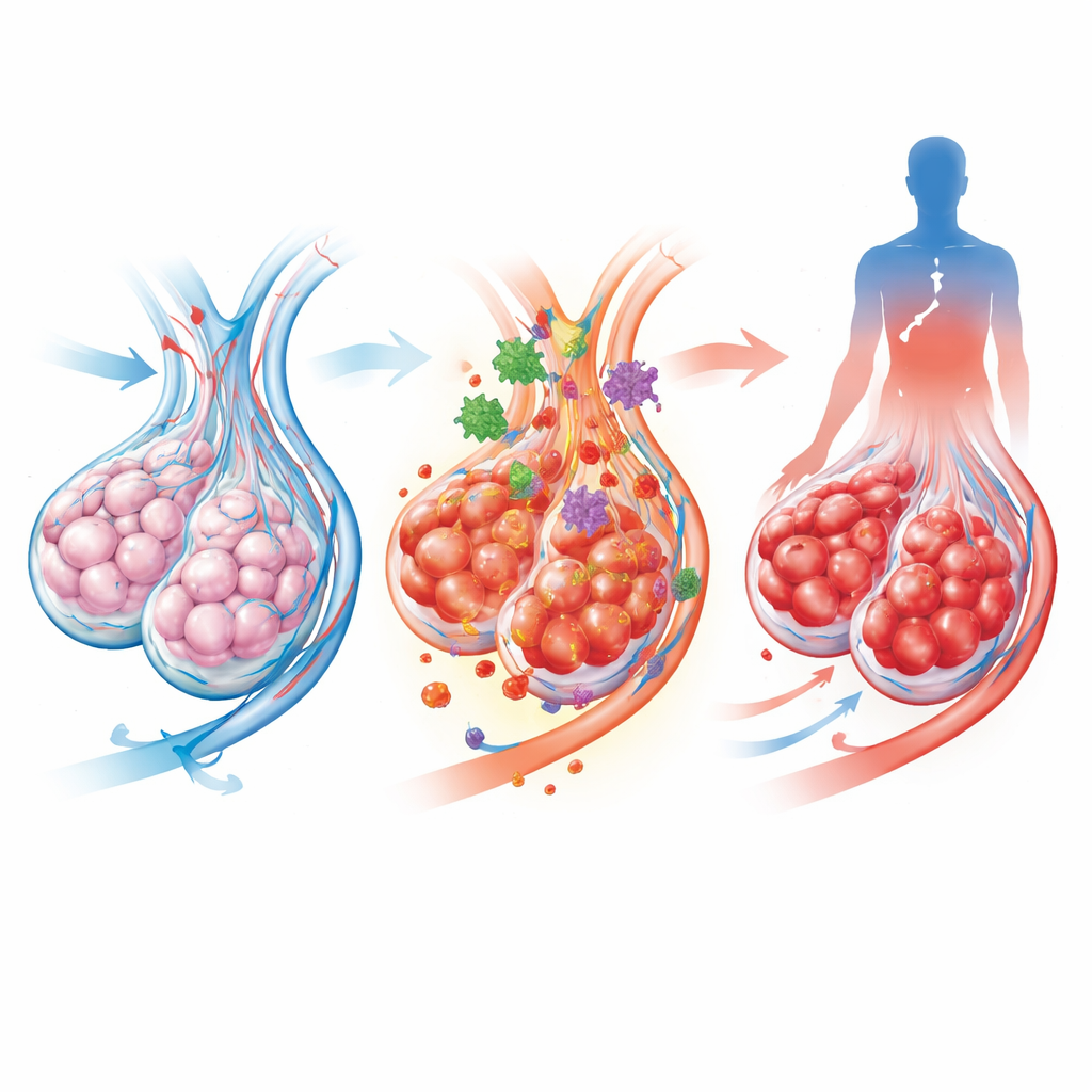
Who Survived and What Predicted Risk
Despite the severity of the condition, in-hospital mortality in this group was 12 percent, lower than many earlier reports. When the team looked for factors linked to death, several stood out in simple one-to-one comparisons: very low oxygen levels on arrival, chest pain, the need for blood transfusions, and finding bacteria in lung samples all pointed toward worse outcomes. Interestingly, patients whose bleeding stemmed from autoimmune disease tended to fare better than those with non-immune causes, likely because prompt immune-targeted therapy can reverse the underlying attack. In a more rigorous combined analysis, one factor remained independently tied to death: hypoxemia, meaning markedly reduced oxygen in the blood at admission. This suggests that how hard the lungs are struggling at the very start of care may matter more than many other lab or demographic details.
What This Means for Patients and Care
For lay readers, the message is both sobering and hopeful. Diffuse alveolar hemorrhage is rare, dramatic, and dangerous, but careful diagnosis and early, aggressive treatment—especially when the cause is an immune disease—can keep many patients alive. The study from Bogotá underscores that doctors should think quickly about this diagnosis when a breathless, anemic patient shows diffuse hazy spots on lung imaging, and they should pay close attention to oxygen levels as a simple, powerful gauge of risk. By recognizing the condition early, clarifying whether the immune system is to blame, and treating both the bleeding and its cause, clinicians can improve the chances that damaged lungs recover before permanent harm is done.
Citation: Lutz, J.R., Martínez-Brocato, E., Segura-Martínez, S.V. et al. Clinical, radiological and laboratory features and outcomes of diffuse alveolar hemorrhage. Sci Rep 16, 8643 (2026). https://doi.org/10.1038/s41598-026-41864-x
Keywords: diffuse alveolar hemorrhage, autoimmune lung disease, pulmonary vasculitis, hypoxemia, bronchoalveolar lavage