Clear Sky Science · en
Diagnostic performance of artificial intelligence for detecting peritoneal and small bowel dissemination in epithelial ovarian cancer using preoperative contrast-enhanced CT imaging
Why spotting hidden spread matters
For women with ovarian cancer, what doctors see inside the abdomen before surgery can make the difference between a curable operation and a major procedure that still leaves tumor behind. Tiny cancer deposits that coat the lining of the abdomen and the surface of the small intestine are especially troublesome. They are hard to see on routine scans yet can prevent surgeons from safely removing all disease. This study explores whether artificial intelligence (AI) can read standard contrast-enhanced CT scans more effectively than the human eye to find this hidden spread and guide safer, smarter treatment plans.
How ovarian cancer quietly spreads
Epithelial ovarian cancer often goes unnoticed until it has seeded the abdomen with small tumor spots. These deposits can carpet the inner lining of the belly and the surfaces of organs, including the delicate loops of the small intestine. When the tumor load is very high or involves critical areas, surgeons may not be able to remove all visible disease, even with extensive operations. Yet survival is best when no tumor is left behind. Today, doctors rely on CT scans to estimate how far the cancer has spread, but detecting tiny spots—especially on the moving, folded small bowel—is difficult. In some cases, surgeons must perform an exploratory operation just to decide whether full tumor removal is possible.
Teaching computers to read the scans
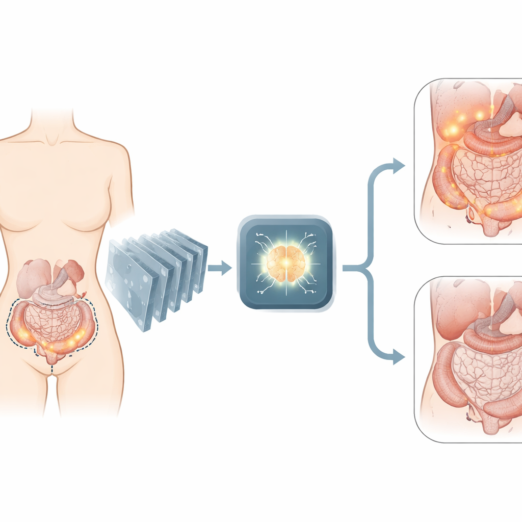
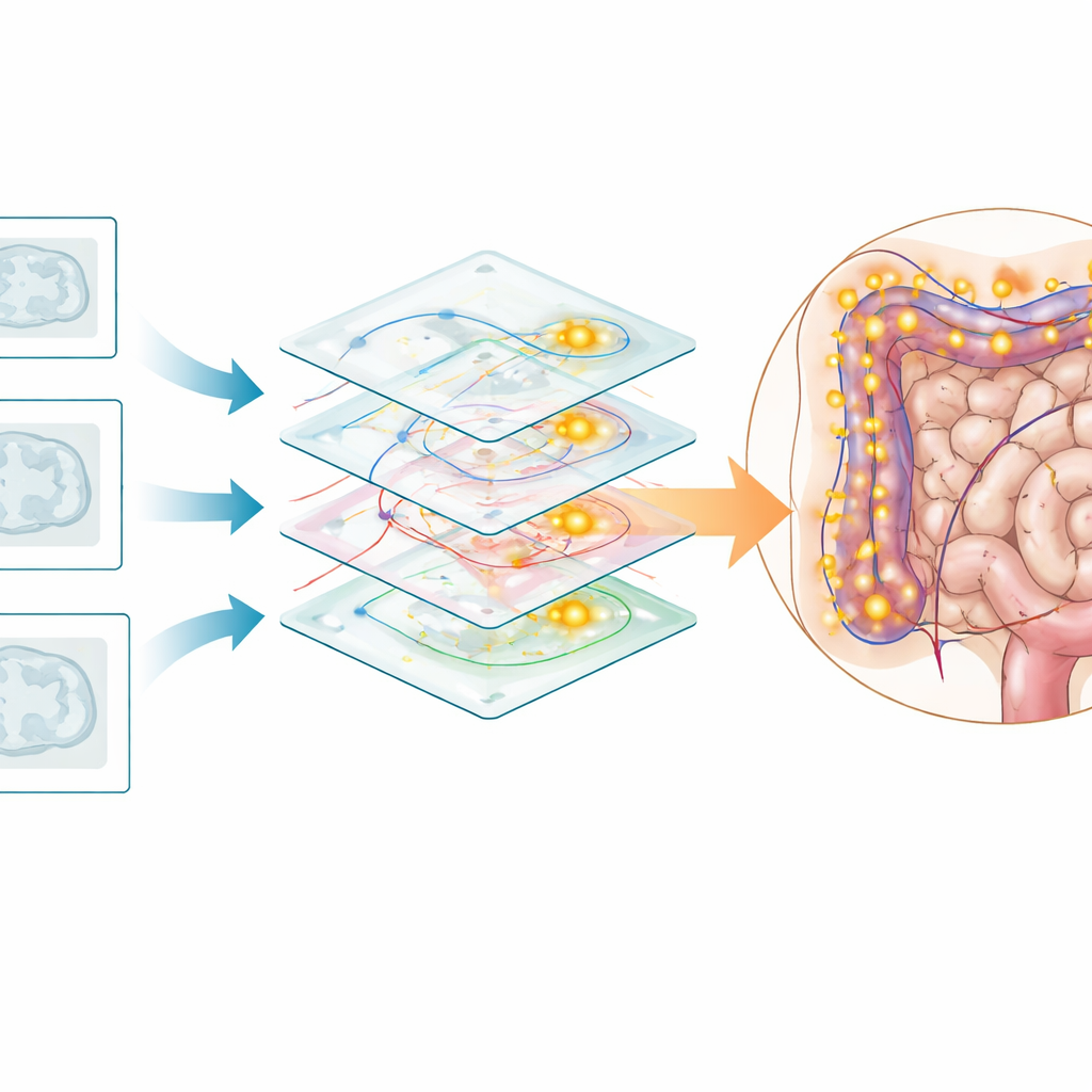
The research team gathered contrast-enhanced CT scans from 227 women treated for ovarian, fallopian tube, or primary peritoneal cancer at two hospitals, yielding 254 scan sets. For each patient, they knew from surgery whether cancer had spread on the abdominal lining and on the small bowel. Using these real-world results as the “answer key,” the authors trained two deep learning systems. One, the P-Model, learned to judge whether the peritoneal surfaces in general carried tumor deposits. The other, the SB-Model, focused specifically on spread involving the small bowel. Both systems were built on a compact neural network design well suited to medical images and were trained and tested repeatedly in different patient groupings to check how stable their performance was.
How well the AI actually performed
When tested on unseen scans, the AI showed promising accuracy. For overall peritoneal spread, the P-Model correctly identified cases about three-quarters of the time, with sensitivity around two-thirds and specificity above four-fifths. In practical terms, it missed some positive cases but produced relatively few false alarms. The small bowel system did even better: the SB-Model correctly classified more than four out of five patients, catching about 86 percent of those with small bowel involvement while still correctly reassuring roughly 77 percent of those without it. This level of sensitivity clearly exceeds the modest performance reported for standard CT reading in earlier studies, where tiny intestinal deposits often went undetected.
When the computer struggled
The researchers also examined situations where the AI performed poorly, defined as being correct in no more than a quarter of its decisions for a given patient. Interestingly, human radiologists also had difficulty in many of these same cases, suggesting that some scan patterns are inherently hard to interpret. The team found that the AI tended to overestimate spread when large amounts of fluid filled the abdomen and tumor markers in the blood were very high, and it sometimes underestimated disease when the tumor burden and fluid volume were low. This pattern hints that the system may have learned to rely heavily on visual cues such as fluid around the intestines, which do not always correspond neatly to how much tumor is actually present.
What this could mean for patient care
Despite its limitations, the study shows that an AI assistant reading ordinary CT scans can meaningfully improve detection of subtle tumor spread, particularly on the small intestine, where radiologists currently have the least confidence. If further tested and refined, such tools could help doctors decide more accurately who is likely to benefit from an aggressive first operation and who might be better served by chemotherapy before surgery. The authors emphasize that AI will not replace expert judgment or exploratory surgery in all cases, but it could become a powerful extra set of eyes, turning existing imaging into a more reliable map for complex cancer surgery.
Citation: Kim, R., Seki, T., Noda, K. et al. Diagnostic performance of artificial intelligence for detecting peritoneal and small bowel dissemination in epithelial ovarian cancer using preoperative contrast-enhanced CT imaging. Sci Rep 16, 8739 (2026). https://doi.org/10.1038/s41598-026-41728-4
Keywords: ovarian cancer, artificial intelligence, CT imaging, peritoneal metastasis, small bowel dissemination