Clear Sky Science · en
Brain connectivity and its relation to cognitive function in patients with post-COVID 19 condition after mild infection
Why this matters for everyday life
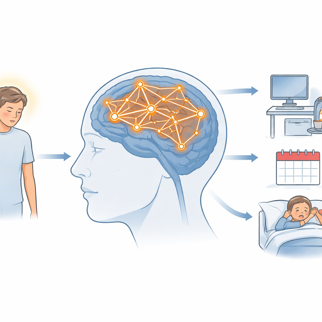
Many people who had only a mild case of COVID-19 still struggle months or even years later with brain fog, trouble concentrating, and exhausting fatigue. Because ordinary brain scans often look normal, both patients and clinicians are left wondering: is there really something different in the brain, or are the symptoms only subjective? This study set out to look under the hood of the resting brain, using a sensitive MRI technique to see whether the “idling” brain networks of people with post-COVID-19 condition (PCC) work differently from those of people without lingering symptoms.
Who the researchers looked at
The team studied 22 adults in Sweden who had a confirmed mild SARS-CoV-2 infection, were never hospitalized, but later developed long-lasting cognitive problems and fatigue that interfered with work and daily life. On average, their symptoms had lasted for almost three years. They were compared with 19 volunteers of similar age and sex who did not report ongoing post-COVID problems. Everyone completed detailed questionnaires on fatigue, anxiety, and depression, plus a battery of cognitive tests focusing on attention, memory, and thinking speed. All participants then underwent advanced brain MRI, including a method that measures how different brain regions naturally “talk” to each other while a person rests quietly in the scanner.
Peeking at the brain’s idle mode
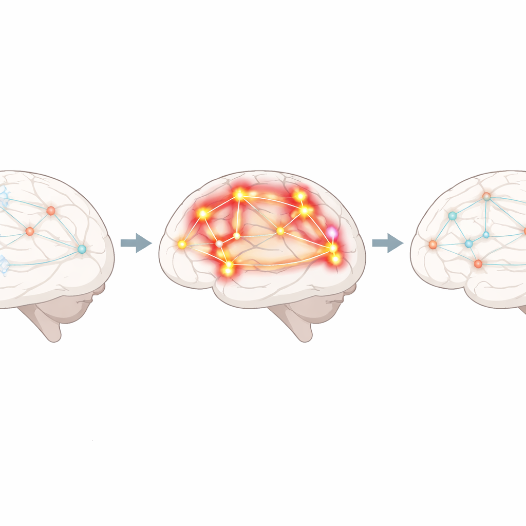
Rather than asking people to perform a complex task, the researchers recorded “resting-state” brain activity, when participants simply stared at a fixed point and let their minds wander. This reveals large-scale networks that turn on and off together. One of the most important of these is the default mode network, a set of regions active when we are awake but inwardly focused—daydreaming, recalling memories, or reflecting on ourselves. Using mathematical tools to break down the MRI signals into independent networks, the team compared how strongly each person’s brain regions were linked within these networks, both before and after a simple sustained-attention task performed in the scanner.
What was different in people with long COVID
The key finding was that, before the attention task, people with PCC showed stronger connections within parts of the default mode network than the comparison group. This heightened “chatter” appeared in areas involved in self-focused thought and visual processing, including the precuneus (a central hub for integrating information), the insula (important for internal bodily awareness), the cerebellum, and regions that help us process complex visual scenes and faces. Interestingly, when the researchers looked at brain activity after the attention task, these differences faded, and the networks of patients and controls looked more similar. At the same time, standard cognitive test scores did not differ significantly between groups, even though the PCC group tended to perform a bit more slowly on visually demanding tasks and reported far more fatigue and mild depressive symptoms.
Clues, but not yet a full explanation
Despite the clear differences in how the default mode network was wired at rest, the study did not find direct links between these brain changes and how people scored on memory and attention tests, or how severely they rated their fatigue, anxiety, and depression. There were also no signs that body weight influenced these connectivity patterns. The authors suggest that the small sample size and the high educational background of many participants may have made it harder to detect subtle drops in performance, because some individuals might still score in the “normal” range despite having declined from their own premorbid level. They also note that elevated connectivity in regions tied to vision and internal awareness echoes other work showing eye-movement problems and visual complaints in PCC, hinting that disrupted communication in these networks could underlie some symptoms.
What this means going forward
For people living with long COVID after a mild infection, this study provides objective evidence that their brains can function differently, even years after the initial illness and even when routine scans appear normal. The altered activity in the brain’s default “idle” network suggests that the resting brain may not be as restorative as it should be, potentially contributing to persistent fogginess and tiredness. Although the study cannot yet tie these changes neatly to specific symptoms or predict who will recover, it underscores that PCC is associated with subtle but lasting shifts in brain function. Larger, longer-term studies will be needed to confirm these patterns and, ultimately, to design targeted rehabilitation strategies that help the brain’s networks return to a healthier balance.
Citation: Hedström, S., Stenberg, J., Borg, K. et al. Brain connectivity and its relation to cognitive function in patients with post-COVID 19 condition after mild infection. Sci Rep 16, 8152 (2026). https://doi.org/10.1038/s41598-026-41665-2
Keywords: long COVID, brain networks, resting-state fMRI, cognitive fatigue, default mode network