Clear Sky Science · en
Evaluation of S-Value in relation to cochlear anatomy in pediatric cochlear implant users
Why this research matters for children with severe hearing loss
Cochlear implants have transformed life for many children born with severe hearing loss, allowing them to develop spoken language and engage more fully with the hearing world. But to get the best possible results, surgeons must thread a delicate electrode array deep into a tiny snail-shaped structure in the inner ear—the cochlea—without causing damage. This study asks a very practical question: can a lesser-known feature of cochlear shape, called the “S-value,” help doctors better plan surgery and keep the implant’s tiny electrodes in the right place?
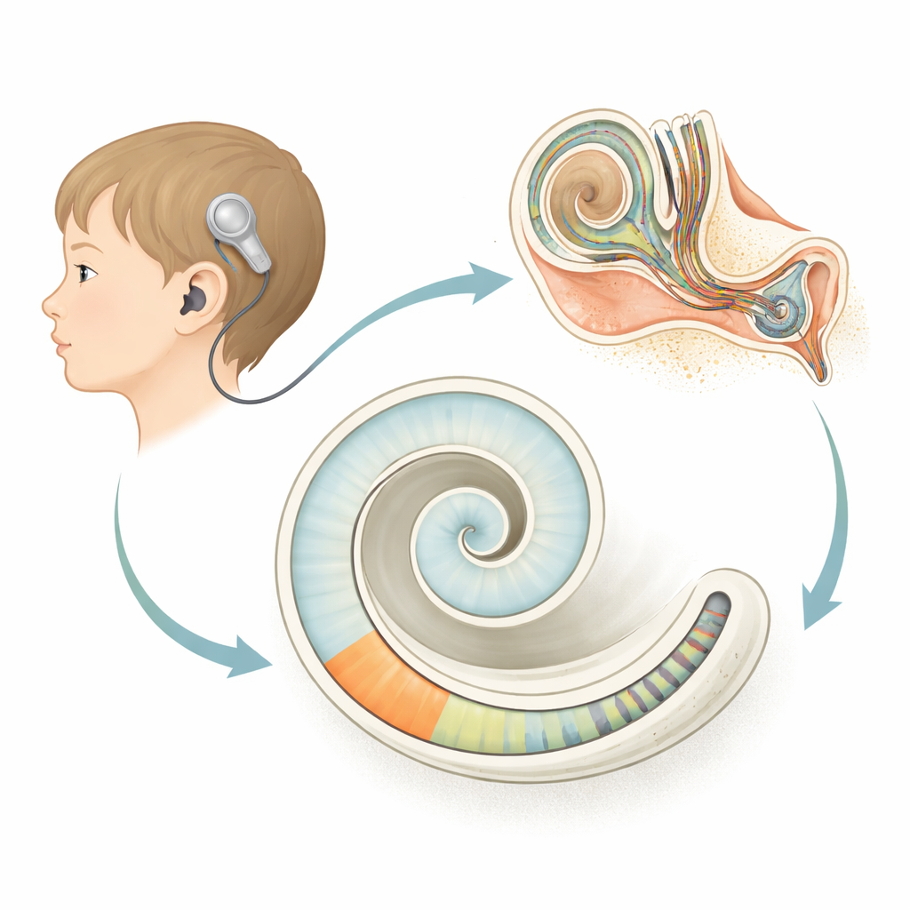
Looking closely at the inner ear’s spiral shape
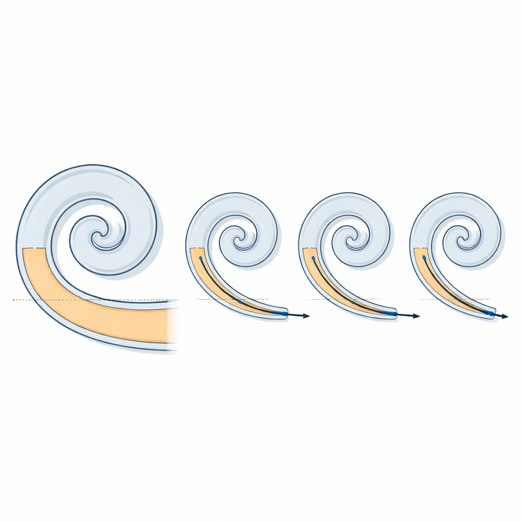
The cochlea is a coiled tube, and its size and shape differ from person to person, even when there are no obvious malformations. Before surgery, doctors routinely use CT scans and planning software to measure basic dimensions such as how long the cochlear tube is and how wide its base is. These measurements help them choose an electrode array that is long enough to cover the hearing region but not so long that it risks bending or causing trauma. The S-value adds another layer of detail: it is the length of the first straight stretch of the cochlear spiral, just as the electrode begins its journey. That straight portion is also the first place where the electrode touches the outer wall of the cochlea, so its length might influence how smoothly the array advances inside.
How the researchers studied children’s cochleae
The authors reviewed high-resolution CT scans from 18 ears in pediatric cochlear implant users treated at a single center. All children had normal inner-ear anatomy and received the same type of straight lateral-wall electrode array (FLEX28) inserted through the round window of the cochlea. Using specialized planning software, two independent reviewers measured the main cochlear dimensions and the S-value. After surgery, the same software was used to examine follow-up CT scans and calculate how far around the spiral each individual electrode contact had traveled—its “angular insertion depth,” which indicates how much of the cochlea the array ultimately covered.
What the team discovered about cochlear shape
Although the overall cochlear duct length in these children was remarkably similar, the size of the straight basal segment showed more variation. The researchers found that the S-value was strongly linked to one key dimension: the diameter of the basal turn (called the A-value). Ears with a larger basal diameter tended to have a longer straight section. In contrast, cochlear width (the B-value) showed only a moderate and statistically non-significant relationship with the S-value. This suggests that, at least in this group of children, the A-value is a better guide to how long the initial straight stretch of the cochlea will be than the overall width of the structure.
Electrode placement despite anatomical differences
The team then asked whether these differences in the straight segment translated into differences in how deeply the electrodes were inserted. Because the same electrode model was used and the total cochlear length was nearly identical across patients, this provided a clean test. They observed only modest, non-significant links between the S-value and insertion angles at each electrode contact. In practical terms, the lateral-wall electrode reached very similar depths around the spiral in all children, regardless of the small differences in S-value. Cochlear coverage—the proportion of the cochlea spanned by the array—was consistently high (around three-quarters of the duct) with very little variation.
What this means for future cochlear implant planning
This study shows that the straight portion of the cochlear base is a reliable anatomical feature that scales with the basal diameter but does not, by itself, strongly alter how deeply a standard lateral-wall array reaches in cochleae of similar overall length. For clinicians, incorporating the S-value into preoperative imaging may still be valuable: it offers a more complete picture of cochlear shape and could help anticipate difficult insertions or guide electrode choice in cases with more unusual anatomy. For families, the main message is reassuring—the particular electrode used in this study tended to sit in a consistent, well-covered position, even when children’s cochleae were not exactly the same shape. Larger, prospective studies comparing different electrode designs will be needed to confirm how best to use the S-value to further personalize cochlear implant surgery and potentially reduce the risk of partial insertions.
Citation: Salamah, M.A., Abdelsamad, Y., Alwhaibi, B. et al. Evaluation of S-Value in relation to cochlear anatomy in pediatric cochlear implant users. Sci Rep 16, 8686 (2026). https://doi.org/10.1038/s41598-026-41504-4
Keywords: cochlear implants, pediatric hearing loss, inner ear anatomy, electrode insertion, medical imaging