Clear Sky Science · en
Estradiol and progesterone regulate secretion and metabolite content of extracellular vesicles from immortalized bovine uterine epithelial cells
How early life depends on hidden helpers
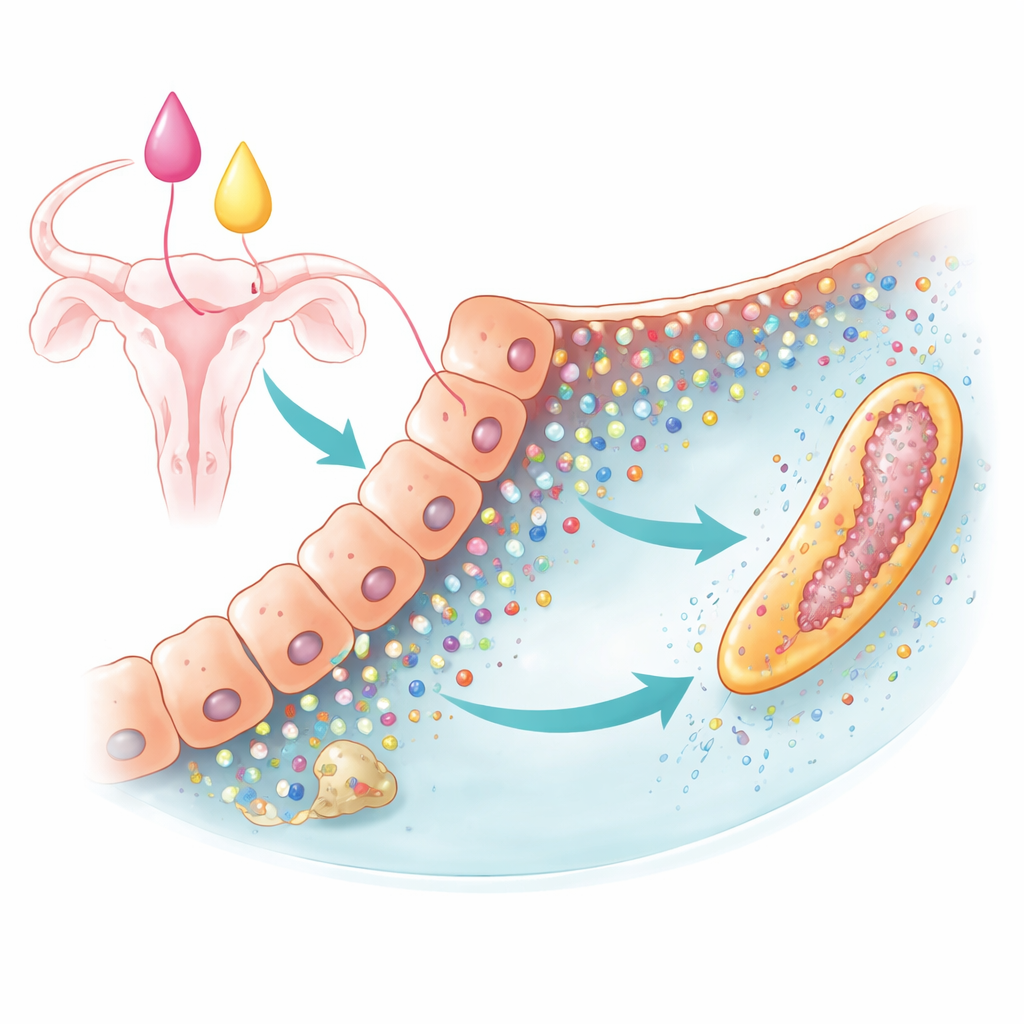
Long before a calf has a heartbeat, its tiny embryo is already busy dividing, stretching, and reshaping itself inside the cow’s uterus. During this delicate window, the embryo cannot feed itself in the usual way and instead depends entirely on secretions from the uterine lining. This study asks a new question about that early support system: do small, membrane-bound particles released by uterine cells act as tiny "care packages" of nutrients, and how do the cow’s own hormones change what is packed inside them?
Tiny packages in the womb
Cells of the uterine lining constantly release minuscule, bubble-like particles known as extracellular vesicles. Although invisible to the naked eye, these vesicles can carry important cargo from one cell to another. In cows, vesicles from the uterus are known to contain genetic material and proteins that influence embryo growth. Yet the basic nutrient molecules—the metabolites—that they might deliver had not been studied. The authors used an immortalized line of bovine uterine epithelial cells, a long-lived lab-grown stand-in for the real uterine lining, to explore what these vesicles look like and what they carry.
Building a lab model of the uterine environment
The team first confirmed that their cell line released genuine extracellular vesicles. They collected the culture fluid from these cells and spun it at very high speeds to pellet the particles. Using particle tracking instruments, they found abundant vesicles mostly in the 100–300 nanometer range, matching the size of known vesicle types. Electron microscopy revealed the classic cup-shaped appearance seen for vesicles inside actual cow uterine tissue. Protein markers that are typical for vesicles were present, while markers of broken cells were absent. Together, these checks showed that the lab-grown cells produce vesicles closely resembling those in the living uterus, making them a solid model for further study.
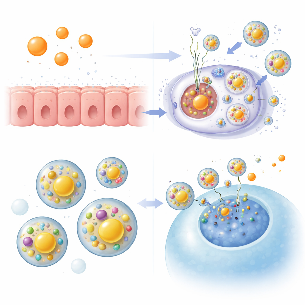
Hormones that reshape the message
Cows’ ovaries produce two key sex hormones, estradiol and progesterone, which naturally rise and fall through the reproductive cycle. The researchers treated the uterine cell line with each hormone and measured vesicle release. Both hormones clearly boosted the number of vesicles the cells secreted, without changing their size. Next, the team used broad, untargeted chemical profiling to catalog the small molecules inside these vesicles. They detected 83 different metabolites, with many related to amino acids, plus some linked to sugars and fats. This mix suggests that vesicles may act as multi-ingredient nutrient packets for the developing embryo, potentially shielding their contents from degrading enzymes in the uterine fluid.
Shifting the nutrient mix
When the cells were exposed to estradiol or progesterone, the chemical cargo inside vesicles shifted in distinct ways. An overall analysis of all detected molecules showed that pathways tied to amino acid use were strongly represented. But when the team zoomed in on the metabolites that actually changed in response to hormones, a different pattern emerged: many of the altered molecules were connected to fat metabolism. Estradiol reduced several fatty acids and related compounds while increasing a shorter-chain fatty acid, and progesterone changed a separate set of fat-like molecules and tended to raise lactic acid. These shifts point to hormone-controlled tuning of lipid components that could help build new cell membranes as the embryo rapidly elongates and the outer cell layer expands.
What this means for early pregnancy
The study concludes that this uterine cell model reliably produces extracellular vesicles whose contents are shaped by estradiol and progesterone. These vesicles carry a variety of amino acid, sugar, and especially lipid-related metabolites, and the two hormones both increase vesicle release while reshaping their nutrient makeup. For a lay reader, the take-home message is that, even before a placenta forms, the mother’s hormones appear to fine-tune tiny nutrient parcels sent from the uterine wall to the embryo. Although the work was done in cultured cells and did not directly track nutrient transfer into embryos, it provides a mechanistic glimpse of how hormonal cycles may prepare the womb to fuel early growth and may guide future efforts to understand fertility and early pregnancy loss in cattle and, by extension, other mammals.
Citation: Sandoval, K., Berg, M.D., Southey, B. et al. Estradiol and progesterone regulate secretion and metabolite content of extracellular vesicles from immortalized bovine uterine epithelial cells. Sci Rep 16, 10249 (2026). https://doi.org/10.1038/s41598-026-41146-6
Keywords: extracellular vesicles, uterine environment, embryo nutrition, reproductive hormones, bovine pregnancy