Clear Sky Science · en
Efficient cosine-windowed cross-correlation for intermediate deformable image registration
Helping Doctors Compare Medical Images More Reliably
Modern medicine often relies on comparing medical scans taken at different times or from different people—for example, to see how a tumor responds to treatment or to build atlases of the brain. But lining up these images so that the same anatomical point appears in the same place is surprisingly hard. This paper introduces a new computational step that makes such alignments faster and more reliable, especially when the anatomy has changed a lot between scans.
Why Lining Up Medical Scans Is So Tricky
When computers align two images, they usually start by correcting big, simple differences such as shifts, rotations, and overall size changes—this is called affine registration. However, real human anatomy bends, grows, and shifts in complex ways that can’t be captured by simple stretching and rotating. Detailed “deformable” registration methods try to fix this by allowing each tiny region to move separately, but they often rely on very local image details. If the changes between scans are large—such as before and after surgery or across different patients—these methods can get stuck in a wrong solution or take a very long time to converge.
A Middle Step Between Coarse and Fine Alignment
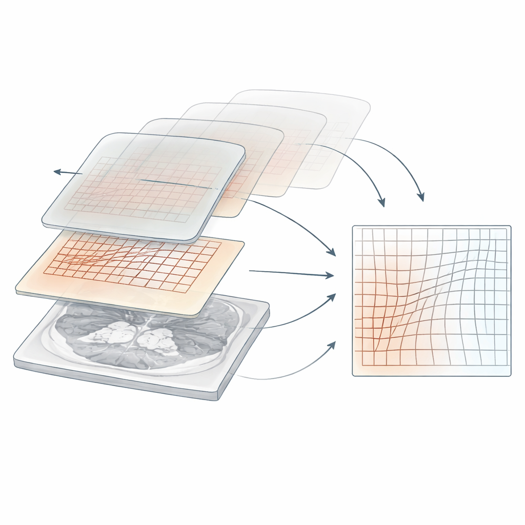
The author proposes an “intermediate deformable image registration” (IDIR) method designed to sit between the coarse affine step and the very fine deformable step. Instead of looking at either the whole image at once or only tiny neighborhoods, IDIR uses very large overlapping windows that slide across the image. Within each window, it estimates how much one image must be shifted locally to best match the other. By choosing a smooth, cosine-shaped window and carefully combining information from all positions, the method produces a smoothly varying map of how each location should move. This map corrects large deformations in just a few iterations, giving later, more detailed methods a much better starting point.
Using Sound-Inspired Math for Faster Matching
Under the hood, the method relies on cross-correlation—a way of measuring how similar two signals are as one is shifted relative to the other. This idea is commonly used in signal processing, such as audio and radar. To keep the computation practical for large images and 3D volumes, the author uses the fast Fourier transform (FFT), which dramatically speeds up correlation calculations. A key innovation is applying cosine-shaped windows to the images before correlating them, and then cleverly expanding the math so that many local shifts can be computed at once instead of one by one. This reduces the computational cost from something that would be prohibitive for real data to something that runs in seconds to minutes on typical hardware.
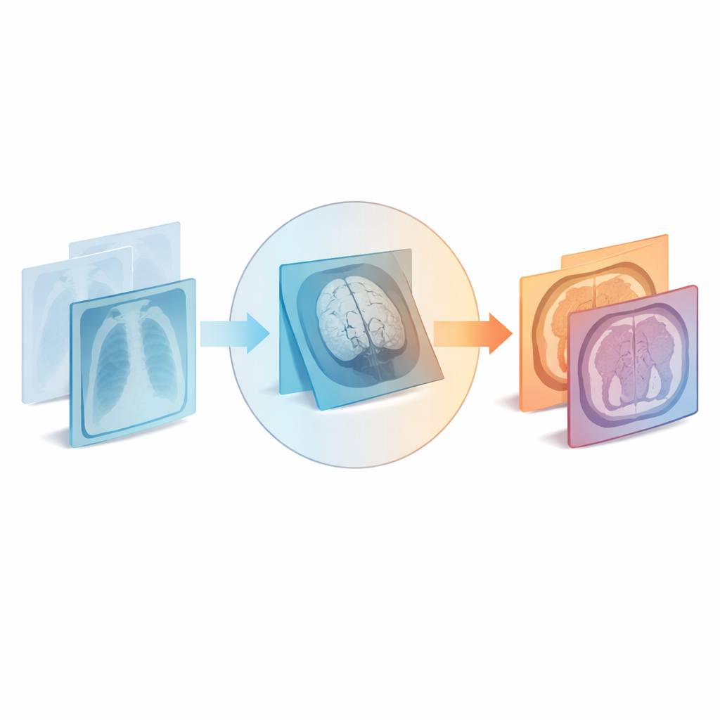
Testing on X-Rays, Brain Scans, and Abdominal CT
The method was tested on three very different kinds of medical images: 2D X-rays of jaws and feet taken before and after surgery, 3D MRI scans of developing fetal brains at different weeks of gestation, and 3D CT scans of the abdomen from different patients. In the X-ray experiments, the new approach quickly captured large surgical changes within a few iterations, producing smooth displacement fields without needing extra smoothing tricks. On the fetal brain MRI, it substantially improved the overlap of labeled brain regions and outperformed a standard deformable algorithm when both were run from scratch. When that same standard method was initialized with the new IDIR result, the alignment improved further. In abdominal CT, the new method again improved organ overlap scores and, when combined with an existing deformable method, beat either one alone for every organ tested.
What This Means for Future Medical Imaging
For non-experts, the bottom line is that this work offers a new way to “pre-align” medical images when anatomy differs a lot between scans. By efficiently correcting large-scale shape differences without requiring any training data or tuning to a specific organ, the proposed IDIR method can make established deformable registration tools more accurate and faster to converge. It is not meant to replace detailed registration entirely but to give those methods a strong head start. Because it is general-purpose and works across X-ray, MRI, and CT, it could be widely useful in research studies and, potentially, in clinical workflows where reliable comparison of medical images is crucial.
Citation: Aganj, I. Efficient cosine-windowed cross-correlation for intermediate deformable image registration. Sci Rep 16, 8629 (2026). https://doi.org/10.1038/s41598-026-40961-1
Keywords: medical image registration, deformable registration, Fourier-based alignment, cross-correlation, medical imaging analysis