Clear Sky Science · en
Integrated transcriptomic analysis of the temporal cortex identifies CRH and GAD2 as neuropathological markers and reveals altered immune microenvironment in Alzheimer’s disease
Why this matters for brain health
Alzheimer’s disease slowly robs people of memory and independence, yet we still struggle to diagnose it early and to understand why the brain’s own defenses sometimes make things worse. This study focuses on a key brain region for memory, the temporal cortex, to search for molecular warning signs in nerve cells and to map how the brain’s immune landscape changes in Alzheimer’s. By combining large genetic datasets with laboratory tests, the authors spotlight two genes, CRH and GAD2, as promising markers of damaged nerve circuits and reveal a striking reshaping of immune cells inside the diseased brain.
Looking inside a memory hub of the brain
The temporal cortex helps us recognize faces, understand language, and store everyday memories. It is also one of the regions most heavily damaged in Alzheimer’s disease. The researchers gathered gene activity data from hundreds of postmortem temporal cortex samples taken from people with and without Alzheimer’s. Because these data came from different studies and laboratories, the team used statistical methods to correct for technical differences and then looked for genes that consistently showed higher or lower activity in Alzheimer’s brains. They found 98 such genes, most of which were turned down rather than up, hinting at a broad loss of normal nerve-cell function.
What the genes reveal about failing brain circuits
When the team examined what these 98 genes normally do, a clear picture emerged. Many are involved in communication between nerve cells, the release of chemical messengers, and processes linked to learning and memory. Pathways related to the calming brain messenger GABA, hormone signaling, and other brain chemicals were especially affected. This pattern supports the idea that Alzheimer’s is not only a disease of toxic protein buildup but also a disease of disrupted signaling between nerve cells, which undermines the brain’s ability to process and store information.
Two standout markers in nerve cells
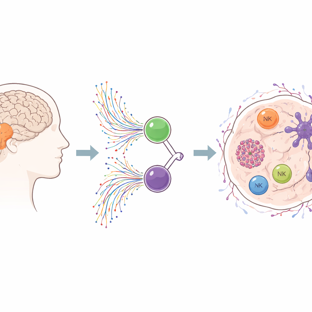
Among the altered genes, two stood out as central players: CRH, which helps coordinate the body’s response to stress and can protect nerve cells, and GAD2, which is essential for making the inhibitory messenger GABA. Both genes were strongly and consistently reduced in the temporal cortex of people with Alzheimer’s across multiple datasets. The authors then confirmed this drop in activity in an independent RNA sequencing study and in new brain samples tested in the lab. When they asked how well these two genes could distinguish Alzheimer’s brains from healthy ones, both showed good diagnostic power on their own, and even better when combined in a simple two-gene model.
A reshaped immune neighborhood in the Alzheimer’s brain
Alzheimer’s is increasingly seen as an immune-related disease, where brain-resident defenders and infiltrating immune cells can either help or harm. Using a computational tool to estimate immune cell types from bulk tissue data, the researchers charted the immune landscape of the temporal cortex. They observed higher levels of cells resembling tissue-repairing macrophages and activated dendritic cells, along with more resting mast cells. At the same time, certain protective or regulating cells—such as plasma cells that make antibodies, regulatory T cells that keep inflammation in check, and activated natural killer cells—were reduced. Together, these shifts point to a chronically inflamed yet imbalanced immune environment in the Alzheimer’s temporal cortex.
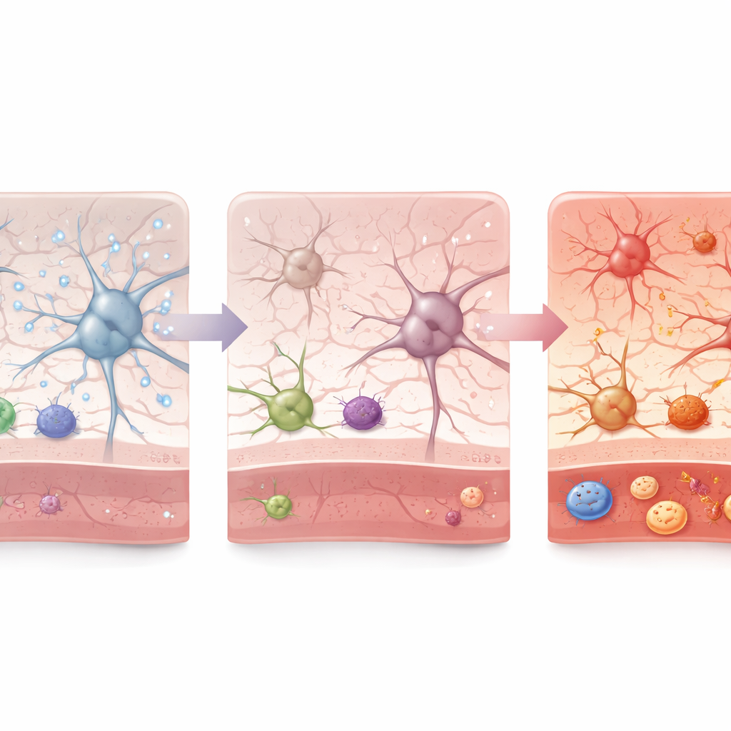
Parallel tracks of nerve and immune damage
One might expect that changes in CRH and GAD2 would closely track the rise or fall of specific immune cells, directly linking nerve-cell stress and immune disruption. Surprisingly, the study did not find strong, simple correlations between the levels of these two genes and the altered immune cell populations. This suggests that nerve-cell gene failure and immune reshaping may be two partly independent dimensions of Alzheimer’s, each driven by complex networks of signals. For non-specialists, the key takeaway is that the disease is not caused by a single culprit. Instead, failing nerve circuits and a misregulated brain immune system appear to march side by side, offering multiple possible targets for earlier diagnosis and for future therapies aimed at both protecting neurons and calming harmful inflammation.
Citation: Liu, P., Huang, C., Lu, L. et al. Integrated transcriptomic analysis of the temporal cortex identifies CRH and GAD2 as neuropathological markers and reveals altered immune microenvironment in Alzheimer’s disease. Sci Rep 16, 10438 (2026). https://doi.org/10.1038/s41598-026-40762-6
Keywords: Alzheimer’s disease, temporal cortex, biomarkers, brain immune cells, gene expression