Clear Sky Science · en
A practical ECG-based model for early identification of acute heart failure following acute myocardial infarction
Why this matters for heart attack survivors
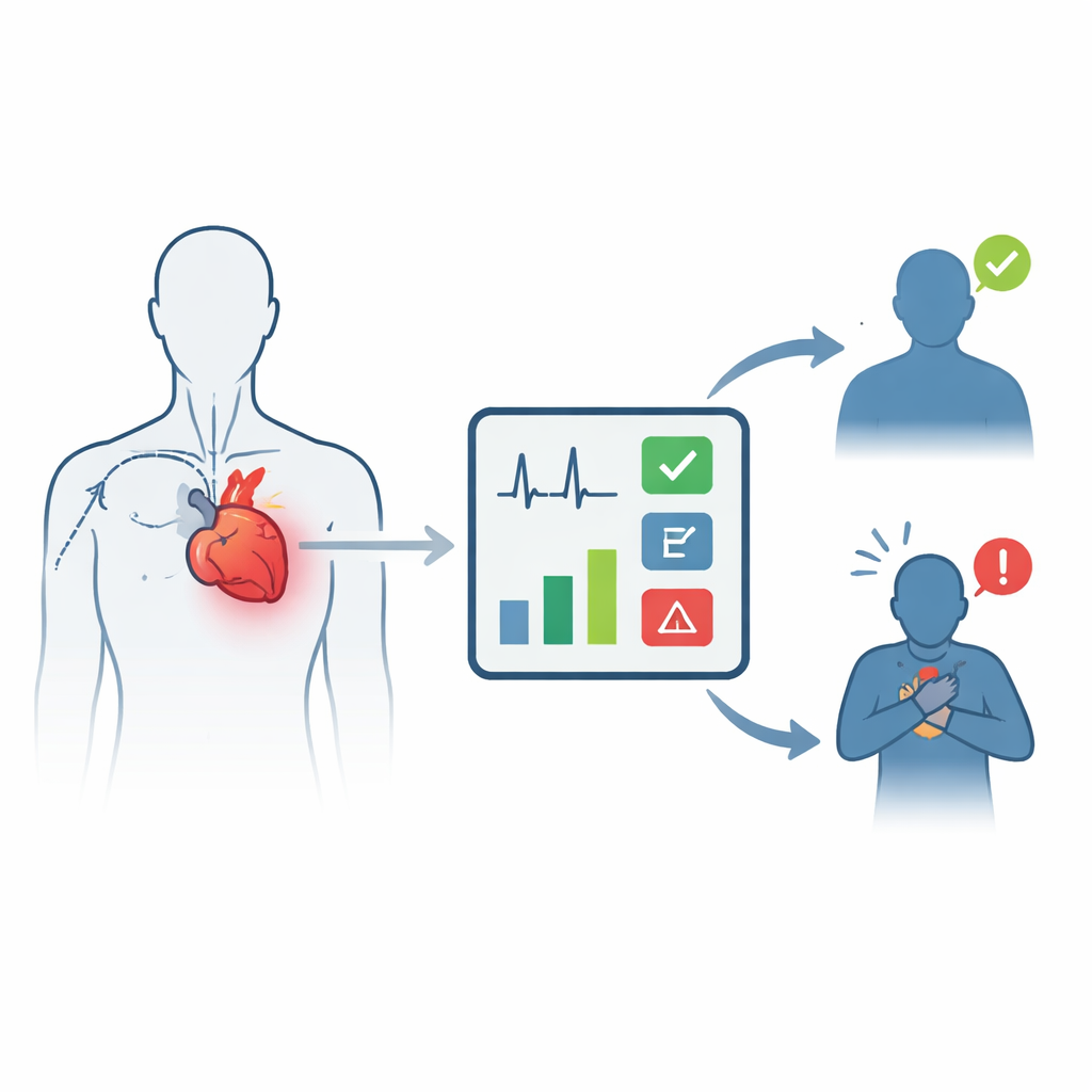
Surviving a heart attack is only the first hurdle. In the days that follow, many patients suddenly slip into acute heart failure, a dangerous state in which the heart cannot pump enough blood to meet the body’s needs. Spotting who is at highest risk early on could save lives, especially in small hospitals with limited equipment. This study asks a simple question with big consequences: can an everyday heart tracing, the electrocardiogram (ECG), be turned into a practical bedside tool to warn doctors which heart attack patients are about to get into serious trouble?
Turning a simple heart tracing into an early warning system
To explore this idea, researchers looked back at 301 people admitted with acute myocardial infarction, the medical term for a heart attack, between 2022 and 2025 at a single hospital in China. Roughly two out of three of these patients developed acute heart failure during their stay. Every patient had a standard 12‑lead ECG recorded on arrival and a routine ultrasound scan of the heart to measure how well the main pumping chamber was working. The team also collected basic clinical information such as age and sex. Instead of relying on costly blood tests or advanced imaging, they focused on ECG patterns that any hospital can measure within minutes.
Finding the most telling signals on the ECG
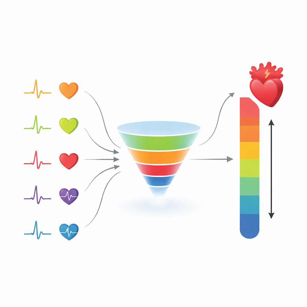
The researchers fed a wide range of ECG measurements into a statistical method designed to filter out weak predictors and highlight the strongest ones. Out of ten candidate ECG features and several clinical factors, six stood out as the most useful for flagging patients likely to develop acute heart failure after a heart attack. These were a prolonged QTc interval (a sign that the heart’s lower chambers take longer than normal to reset between beats), the presence of an abnormal Q wave (often reflecting a larger area of damaged muscle), a resting heart rate above 100 beats per minute, a reduced ejection fraction on ultrasound (indicating weaker pumping), being male, and being between 60 and 75 years of age.
Building a bedside risk score
Using these six factors, the team assembled a visual risk calculator known as a nomogram. In this chart, each factor contributes a certain number of points: for example, a very low ejection fraction adds many points, while a normal heart rate adds few. Adding up the points yields an estimated probability that a given heart attack patient will develop acute heart failure. When the researchers tested how well this tool separated high‑risk from low‑risk patients, it performed better than any single ECG feature alone. The overall accuracy, measured by the area under the receiver operating characteristic curve, was about 0.84, which is considered good for clinical prediction tools. Internal checks using repeated resampling showed that the predicted risks closely matched what actually happened.
What the patterns reveal about disease severity
Beyond predicting who would fail, the study also examined how ECG patterns tracked with the Killip grade, a scale doctors use at the bedside to rate how severe heart failure has become. Patients with higher grades, indicating more severe fluid buildup and breathing problems, tended to have longer QTc intervals, faster heart rates, and were slightly older. Interestingly, certain fine details of the P wave, which reflects the upper chambers of the heart, became shorter and less varied as heart failure worsened, hinting at subtle changes in the heart’s electrical wiring as the disease progresses. These links suggest that the ECG not only warns about risk, but may also mirror how heart failure advances in real time.
Bringing advanced risk prediction to smaller hospitals
Like all retrospective, single‑center studies, this work has limits: the sample was modest, women were under‑represented, and some potentially useful ECG markers were not available. The model still needs to be tested in different hospitals and regions before it can guide everyday care. However, the message for non‑specialist settings is clear. With nothing more than a routine ECG and a basic heart ultrasound, clinicians may soon be able to estimate, at the bedside, which heart attack patients are heading toward acute heart failure and need closer monitoring or more aggressive treatment. If validated more widely, this low‑cost tool could help community and rural hospitals offer earlier, more tailored care to some of their most fragile patients.
Citation: Guo, X., Yan, G., He, H. et al. A practical ECG-based model for early identification of acute heart failure following acute myocardial infarction. Sci Rep 16, 9711 (2026). https://doi.org/10.1038/s41598-026-40600-9
Keywords: acute heart failure, electrocardiogram, acute myocardial infarction, risk prediction model, primary care cardiology