Clear Sky Science · en
Combined proteomics and metabolomics analyses revealed molecular signatures associated with proliferative diabetic retinopathy
Why this matters for people with diabetes
Diabetic eye disease is one of the leading causes of preventable blindness, yet many patients still lose vision even with modern treatments. This study peeks into the “chemical soup” inside the eye to find early warning signs and new treatment targets for the most severe form of the disease, called proliferative diabetic retinopathy. By measuring hundreds of proteins and small molecules at once, the researchers highlight a handful that seem to drive harmful blood vessel growth and inflammation in the diabetic retina.
Looking inside the eye’s inner ocean
The clear gel that fills the eye, known as the vitreous, sits right next to the light-sensing retina and quietly reflects what is happening in that fragile tissue. The team collected undiluted vitreous samples from eight people with advanced proliferative diabetic retinopathy and six non-diabetic patients undergoing surgery for a different, non-inflammatory eye condition. They then used two powerful “omics” methods in parallel: proteomics to catalog proteins, and metabolomics to track small metabolic molecules. Statistical tools were used to see how the diabetic and non-diabetic samples clustered and which components changed most strongly between groups. 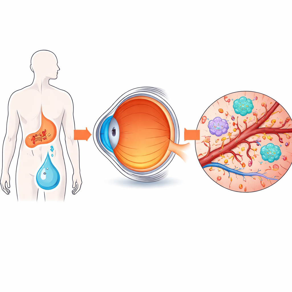
Key troublemakers and missing protectors
The combined analysis revealed 81 proteins and 26 metabolites that differed between diabetic and control eyes. From these, the scientists focused on seven proteins and six metabolites that were most tightly connected. Three proteins stood out. CD5L, an immune-related protein, was higher in diabetic eyes, while CLU (clusterin) and SERPINF1 (which produces a protective factor often called PEDF) were lower. Follow-up tests in patient samples and in a rat model of diabetic retinopathy confirmed this pattern both in the eye fluid and in the retina itself. In simple terms, factors that usually calm inflammation and keep vessels stable were depleted, while a factor that can push cells toward growth and activation was increased.
Energy stress and leaky blood vessels
The metabolite data added another layer to the story. Many of the altered molecules belonged to fatty and organic acid groups, with several linked to energy handling in cells. Creatine, a well-known energy buffer for hard-working tissues like nerves and vessel linings, was reduced. This suggests that retinal cells in diabetes may be running on an unstable energy supply, which can worsen stress and damage. When the researchers combined protein and metabolite results, they saw that these changes were concentrated in pathways that control blood clotting, complement (a part of the immune system), and responses to injury. Together, they point toward a scenario in which chronic high sugar tips the balance toward inflammation, microclots, and leaky, fragile microvessels.
Zooming in on one key signal
To test whether any of the altered proteins actively drive disease-like behavior, the team turned to cell culture. They added extra CD5L to mouse blood vessel cells and watched what happened. With this single change, the cells began to divide faster and move more readily—two basic behaviors required for new vessel sprouts to form. This supports the idea that excess CD5L in diabetic eyes could help fuel the abnormal blood vessel growth that characterizes proliferative diabetic retinopathy. At the same time, the loss of CLU and SERPINF1 likely removes important brakes on inflammation and vessel overgrowth, compounding the problem. 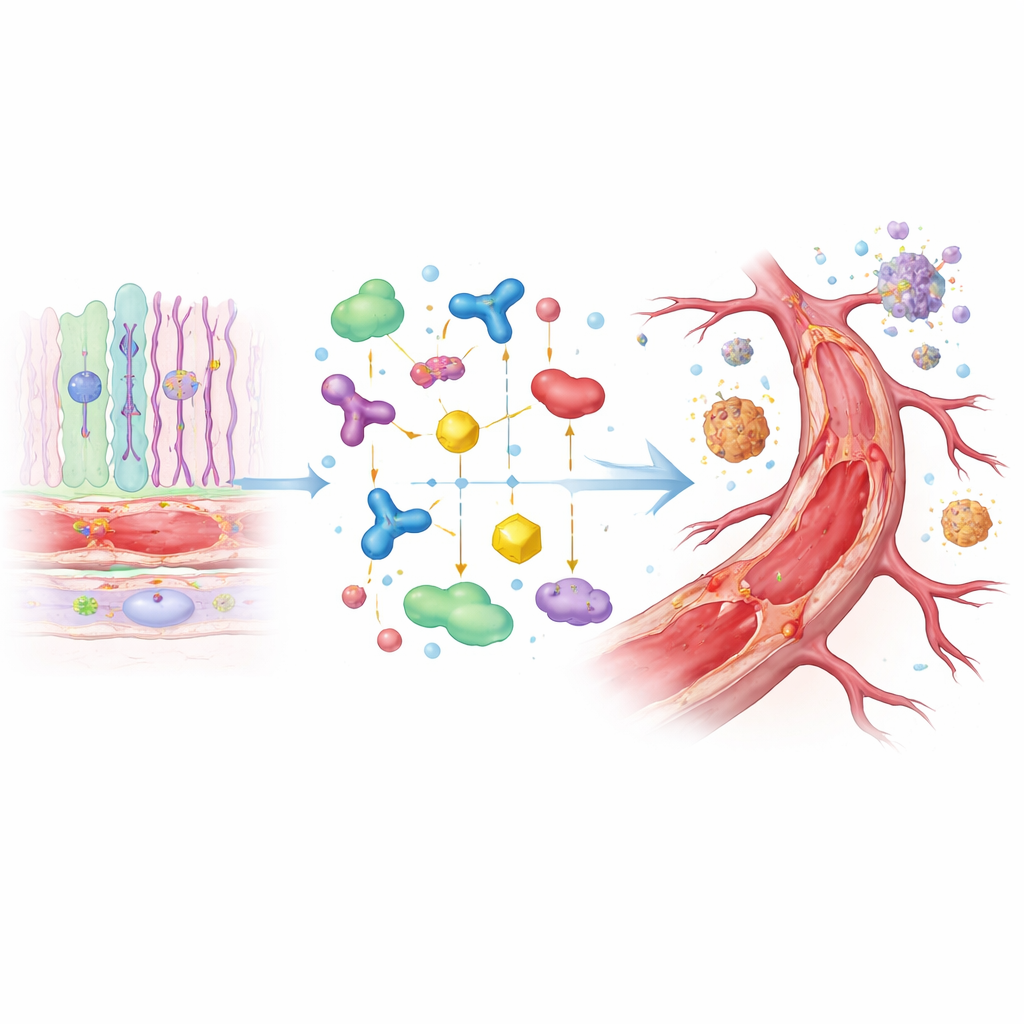
What this could mean for future care
Taken together, the findings sketch a more complete picture of proliferative diabetic retinopathy: not just as a disease of high blood sugar and overactive growth signals like VEGF, but as a coordinated breakdown in immune balance, vessel stability, and cellular energy use. The highlighted proteins and metabolites could serve as biomarkers to flag patients at higher risk before severe damage occurs. They also hint at new treatment avenues, such as drugs that dial down CD5L’s pro-growth influence or restore protective molecules like SERPINF1 and CLU. While the study is small and relies partly on animal models, it offers a roadmap for turning complex molecular data into practical strategies to better protect sight in people living with diabetes.
Citation: Cui, Y., Rao, L., Shen, L. et al. Combined proteomics and metabolomics analyses revealed molecular signatures associated with proliferative diabetic retinopathy. Sci Rep 16, 9755 (2026). https://doi.org/10.1038/s41598-026-40551-1
Keywords: diabetic retinopathy, retinal blood vessels, multi-omics, eye inflammation, biomarkers